
M.S. Shathele
Department of Microbiology and Parasitology, King Faisal University, P.O. Box 1757, Al-Ahsa 31982, Kingdom of Saudi Arabia
A. Fadlelmula
Department of Microbiology and Parasitology, King Faisal University, P.O. Box 1757, Al-Ahsa 31982, Kingdom of Saudi Arabia
Asian Journal of Animal and Veterinary Advances
Year: 2010 | Volume: 5 | Issue: 3 | Page No.: 180-192


ABSTRACT
Three types of common and one commercial antifungal drugs were tested in vitro for assessing their effectiveness against dermatophytic fungi Trichophyton verrucosum using a microdilution assay of the NCCLS (M38-P) standard for filamentous fungi with slight modifications. Four agents of antifungal drugs in different concentrations defined as local mixture [consisting of cupric sulfate and calcium oxide (quicklime) by a mixture proportion of 1:1 in concentration of 80.0 mg mL-1], Hydrogen Peroxide mixture in concentration of 180.0 mg mL-1, 10% Formaldehyde mixture in concentration of 39.0 mg mL-1 and Amphotericin B (AMB) in concentration of 2.0 mg mL-1. The results showed that there is a discrepancy among the antifungal drugs on the impact of dermatophytic fungi Trichophyton verrucosum. The order of effectiveness of four antifungal drugs against dermatophytic fungi was AMB with a Minimum Fungicidal Concentration (MFC) (MFC90% = 2.0 μg mL-1)> 10% Formaldehyde mixture with a concentration of (MFC90% = 3.9 μg mL-1)> Hydrogen Peroxide mixture with a concentration of (MFC90% = 18.0 μg mL-1)> the local mixture with a concentration of (MFC90% = 80.0 μg mL-1). In conclusion, AMB with a Minimum Fungicidal Concentration (MFC) (MFC90% = 2.0 μg mL-1) is the most effective drug against dermatophytic fungi under the climatic condition of AL-Ahsa, Saudi Arabia.
PDF Abstract XML References Citation
How to cite this article
M.S. Shathele and A. Fadlelmula, 2010. In vitro Effectiveness of Some Antifungal Drugs in Treatment of Trichophyton verrucosum; Dermatophytic Fungi. Asian Journal of Animal and Veterinary Advances, 5: 180-192.
DOI: 10.3923/ajava.2010.180.192
URL: https://scialert.net/abstract/?doi=ajava.2010.180.192
DOI: 10.3923/ajava.2010.180.192
URL: https://scialert.net/abstract/?doi=ajava.2010.180.192
INTRODUCTION
The dermatophytes are taxonomically related fungi which have the ability to utilize keratin as a nutrient source i.e., they have a unique enzymatic capacity [keratinase] causing different skin infections referred to as tineas in man or ringworm in man and animals (Macura, 1995; Al-Doory, 1980; Ajello, 1974, 1977). Recently, the number of infections caused by these fungi has increased considerably causing particular concern when they infect immuno-compromised patients where typical manifestations and more severe, extensive lesions can be produced (Macura, 1995; Weitzmann and Summerbell, 1995; Gupta et al., 2001; Berger, 1996; Ray and Gately, 1994). Contagiousness among animal communities, high cost of treatment, difficulty of control and the public health consequences explain their great importance (Chermette et al., 2008). Ring worm is considered one of the most important fungal skin diseases that affect both human and animals alike (Ali-Shtayeh et al., 1988; Del-Rosso, 2000; Tsang et al., 1996; Amer et al., 2006; Wildfeuer et al., 1998; Butty et al., 1995; Nimura et al., 2001). It has been suggested that about 80% of human cases of the disease, especially among children are caused by Microsporum canis from dogs. And the infection is more severe when the disease is transmitted from animals to humans compared to when transmitted from human to human (Grappel, 1981). A wide variety of dermatophytes have been isolated from animals, but a few zoophilic species are responsible for the majority of the cases, viz., Microsporum canis, Trichophyton mentagrophytes, Trichophyton equinum and Trichophyton verrucosum, as also the geophilic species Microsporum gypseum (Hasegawa, 2000; Mahmoudabadi and Zarrin, 2008; Butty et al., 1995). The zoophilic dermatophyte T. verrucosum is associated principally with cattle and camel ringworm (FadlElmula et al., 1994; Lepper, 1972) but it has been reported to infect a wide range of animal hosts together with man (Emmons et al., 1977). In animals, the lesions start with thickening of skin, alopecia and scaliness. They may involve small circular areas or become confluent in extensive areas. The exudates of the inflammatory process glues hair together into thick grey asbestos-like crusts which reveal bleeding ulcerated areas on removal. Ringworm in animals in the Kingdom of Saudi Arabia (KSA) was reported (Gitao et al., 1998). Clinical ringworm in camels was also reported (Al-Hendi et al., 1998). In man, the disease has also been described in the KSA (Venugopal and Venugopal, 1992, 1993). Trichophyton verrucosum, T. mentagrophytes and T. megninii have been regarded as the main fungi causing ringworm in cattle (Quinn et al., 1994; Abou-Gabal et al., 1976; Al-Ani et al., 1995). However, very limited studies on ringworm infection in cattle have been published from Arab countries. Abdullah and Hassan (1995) isolated T. verrucosum, M. fulvum and M. gypseum from surface sediments of the Shatt Al-Arab River of Iraq. Also, T. verrucosum has been isolated from soil in a playground in the Nablus area, West Bank of Jordan (Ali-Shtayeh, 1989). In horses, Trichophyton species and Microsporum species are the main causes of ringworm in Saudi Arabia (Bagy and Abdel-Mallek, 1991). In Sudan Abu-Samra and Ibrahim (1988) found that horses were successfully infected with human isolates of M. canis and T. violaceum. Development of drugs that are effective against fungal cells without being excessively toxic to human cells has been limited because fungal cells are very similar to human cells. A few antifungal agents are available and licensed for use in veterinary practice or human being treatment. The use of systemic drugs is limited to treat man or animal due to their high toxicity and problems of residues in products intended for human consumption (Araujo et al., 2009). Numerous topical agents and several systemic ones are available, but comparison of their in vitro activity against dermatophytes has been hampered by the lack of a well-accepted MIC assay for these fungi (Barchiesi et al., 2001; Espinal-Ingroff, 2001; Hazen, 1998, 2000; Jessup et al., 2000a; Niewerth et al., 1998; Korting et al., 1995; Norris et al., 1999; Wildfeuer et al., 1998). Activity of available antifungal drugs, against strains, representing a wide local isolates of dermatophyte species and following standard procedures, has not yet been investigated. Although, several antimycotic drugs are valuable at present, its use is limited by a number of factors, such as low potency, poor solubility, emergence of resistant strains and drug toxicity. Therefore, there is a distinct need for the discovery of new, safer and more effective antifungal agents. Recently, several groups have adapted the proposed reference method for broth dilution antifungal susceptibility testing of conidium-forming filamentous fungi for developing a more specific assay for dermatophytes (National Committee for Clinical Laboratory Standards, 2002; Fernandez-Torres et al., 2000). Since, the preparation of conidia inoculum is sometimes a challenge with dermatophytes, a microdilution assay appears to be the ideal format (Jessup et al., 2000b; Norris et al., 1999; Fernandez-Torres et al., 2000, 2001). However, assay parameters, such as the temperature, duration, or growth inhibition endpoint, are still the subject of debate (Hofbauer et al., 2002; Jessup et al., 2000a, b; Perea et al., 2001). The NCCLS guidelines are primarily aimed toward susceptibility testing of clinical isolates. The aim of the present study was to establish an NCCLS-compatible assay, which was optimized for our primary purpose of evaluating investigative antifungal agents.
Consequently, the aim of this study has been to evaluate the in vitro activity of four antifungal drugs in common use and of a local mixture agent against local isolates of dermatophytes; T. verrucosum by following the NCCLS guidelines for testing fungi (National Committee for Clinical Laboratory Standards, 2002) (12). The percentage of Minimum Inhibition Concentration (MIC) and Minimum Fungicidal Concentration (MFC) of the antifungal agents are also recorded.
MATERIALS AND METHODS
Dermatophyte Isolates
Dermatophytes belonging to Clinical isolates of T. verrucosum (n = 10) isolates were tested. These clinical isolates (skin, hair) were received from the veterinary hospital during a clinical trial on calves infected with dermatophytic fungi. The reference isolates obtained from Assiut University, Egypt were also tested. The fungi were maintained in sterile distilled water at room temperature (28 to 37°C) and prior to testing were sub-cultured on antimicrobial-agent-free Potato Dextrose Agar (PDA) (sigma) at 37°C for 7 to 15 days to ensure the purity and viability of the inoculum. The strain Paecilomyces variotti (ATCC 36257) was included as quality control. All clinical isolates were identified to species level on the basis of standard biochemical tests, microscopy and colony characteristics. The isolates grown from clinical samples were sub-cultured once to confirm purity and the cultures were maintained in sterile water and on Sabouraud agar slopes.
Medium
The tests were performed in RPMI 1640 medium (Sigma Company USA) with L-glutamine and without sodium bicarbonate buffered at pH 7.0 with (0.165 M = 34.54 g L-1) morpho-line-propane-sulfonic acid (MOPS) (Sigma Company USA). The Media was prepared by following mainly the NCCLS guidelines for testing filamentous fungi (National Committee for Clinical Laboratory Standards, 2002). The Media was filtered by using a sterilized 0.45 mm pore filter and preserved at 4°C temperature until use.
Preparation of Antifungal Drugs Stock Solutions
Amphotericin B (AMB) was obtained from Sigma Company USA; hydrogen peroxide (Sigma Company USA), formaldehyde solution (Sigma Company USA) and a local mixture of copper sulfate + calcium oxide mixed together with concentration of 8% in equal proportion of 1: 1 ratio (commonly used by local farmers and animal breeders). The stock solutions of local mixture were prepared in distilled water at a concentration of 80.0 mg mL-1, then eight concentrations ranging from 0.0008 - 80000 μg mL-1 were selected for experiment. The stock solutions of Hydrogen Peroxide were prepared in distilled water with a concentration of 180 mg mL-1, then eight concentration ranging from 0.0018-180000 μg mL-1 were selected. Similarly, stock solutions of 10% Formaldehyde solution were prepared in distilled water with a concentration of 39 mg mL-1 and then eight concentrations ranging from 0.00039 to 39000 μg mL-1 were selected. The stock solutions of AMB were prepared in 100% dimethyl sulfoxide (DMSO) with a concentration of 2 mg mL-1 (2,000 μg mL-1) then eight concentrations ranging from 0.0002-2000 μg mL-1 were selected. All stock solutions were sterilized by filtration using Sterilized gauze. The stocks solutions were kept at a 4°C until required. All drugs were dissolved and twofold serially diluted as described by the National Committee for Clinical Laboratory Standards (2002) beginning at 100 times the test concentration followed by a further 1:50 dilution in RPMI medium to yield twice the final concentration required for testing. All standard media were purchased from Merck.
Preparation of Fungal Inoculum Suspensions
Mature colonies from 7 to 15 days old cultures grown on PDA at 37°C (Fung-Tomc et al., 1995) were submerged with approximately 10 mL of sterile saline (0.85%) using tip of a sterile Pasteur pipette mycelium and conidia were scraped from the top of the fungal colonies and dispersed in a small volume of Sabouraud 2% dextrose broth (usually 20 mL for 25 plates) using a sterile glass homogenizer. One drop (1%) Tween 20 was added to reduce the surface tension of Fungal cell wall to avoid its sticking to the sides of plates (Espinel-Ingroff et al., 2002). Heavy particles were allowed to settle for 15 to 20 min at room temperature, then the upper suspension was mixed with a vorter mixer for 2 min until it was well homogenized. The turbidity of the supernatants was measured spectrophotometrically at a wavelength of 530 nm and transmission was adjusted to 65-75% for the T. verrucosum in order to have the size of fungal units equal 0.5X 100000-5X 100000 (Espinel-Ingroff et al., 2002). Each suspension was diluted 1:50 in RPMI 1640 to obtain the final test inoculum twice. The inocula corresponding to 10 isolates (T. verrucosum species) were quantified by plating 0.01 mL of a 1:100 dilution of the adjusted inoculum on PDA plates. The plate contents were then incubated at 37°C and were observed daily for the presence of fungal colonies. The viable count was determined as number of CFU per milliliter by serially diluting the stock in 0.86% NaCl and spreading 100 μL plate-1 on the same agar medium as that one used for the inoculum preparation. The final concentration of DMSO was 1% and the inoculum size was 2.9x10⁵ CFU mL-1. Plates were incubated for 4 to 5 days, depending on the growth in control wells without drug, at 37°C for T. verrucosum.
Procedures of Concentration Measurement
The procedures were performed using a broth microdilution method following the National Committee for Clinical Laboratory Standards (2002) guidelines and steps to the test as follows:
Procedures of MIC Measurement
The lowest concentration of the drug inhibitory of fungi growth (approximately 50 and 90% of the growth control). And Determine the Minimum Fungicidal Concentration (MFC); lowest concentration of the drug that are lethal to fungi: (approximately 100% of the growth control). Sterile Microdilution plates with flat-bottom 96 well (Greiner) were set up in accordance with the National Committee for Clinical Laboratory Standards (2002) M38-P method. Aliquots of 100 μL of the 2x drug dilutions were inoculated into the wells with a multichannel pipette. The microplates were stored at 4°C until used. When the susceptibility test was performed, 100 μL of the diluted fungal inoculum suspensions of an exponentially growing culture (2.9x10⁵ CFU mL) was added to each well to bring the drug dilutions to the final test concentrations. The MIC corresponded to the minimum concentration of the compound that caused 99% cell inhibition with respect to the CFU's in a control which contained microbial cultures and sterile distilled water or solvent replacing the test compound.
Test concentrations for local mixture ranged from (0.0008 to 80000 μg mL-1), test concentrations for the Hydrogen Peroxide solution ranged from (0.0018 to 180000 μg mL-1), Test concentrations for Formaldehyde solution ranged from (0.00039 to 39000 μg mL-1) and Test concentrations for AMB ranged from (0.0002 to 2000 μg mL-1). Growth and sterility control wells were included for each isolate tested. The microplates contents were incubated at 37°C and were read by the naked eye after 10 days of incubation, avoiding desiccation. Fungal Growth inhibition was measured visually by the size of the amount of numerical growth, the development of (0-4) according to the NCCLS M38-P reference method as follows:
| • | (0) | : | There is no growth with the naked eye indicating growth rate of 0 and 100% inhibition |
| • | (1) | : | There is a very light growth with estimated growth rate of 25 and 75% inhibition |
| • | (2) | : | There is average growth with estimated growth rate of 50 and 50% inhibition |
| • | (3) | : | There is above-average growth with estimated growth rate of 75 and 25% inhibition |
| • | (4) | : | There is a full growth with estimated growth rate of 100 and 0% inhibition |
Determine the Minimum Fungicidal Concentration (MFC)
Lowest concentration of antifungal drugs that are lethal to fungi WAS PERFORMED After MIC determination, 0.1 mL from the well that had shown no growth at all, which was given the number zero, starting from the last well in which growth was observed up to the highest drug concentration tested, was transferred into glass tubes containing 5 mL of Sabouraud 2% dextrose broth (pH 6.5). and were incubated at 37°C until growth. Plates of growth, has been read to note the extent of growth to determine the MFC as follows: Dish, which grew more than three fungal colonies is not a lowest fungicide concentration. And only Dishs, which grew (1-3) fungal colonies were estimated as the MFC is a killer dish (Fung-Tome et al., 1995).
Data Analysis
The MIC range, the MIC at which 50% of the isolates tested are inhibited (MIC50) and MIC90 are provided.
RESULTS AND DISCUSSION
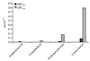
The growth for T. verrucosum could be visualized within 10 days. The Inoculum count ranged from 1.5x10⁵ and 2.2x10⁵ CFU mL-1 with a mean value of 2.9 x10⁵ CFU mL-1 which was used in all tests. There is no consensus concerning the optimal growth inhibition endpoint for MICs (Fernandez-Torres et al., 2000, 2001; Perea et al., 2001). We uniformly adopted a score of 1 as the MIC for all the tested drugs as recommended by Norris et al. (1999). The results of MIC and the in vitro activities of 4 antifungal agents against T. verrucosum species of dermatophytes represented in Table 1. The MIC range for AMB was the narrowest (0.0002-0.2 μg mL-1) and that for Local mixture was the widest (0.008- 80 μg mL-1).
| Table 1: | In vitro activities of 4 antifungal agents against T. verrucosum dermatophyte |
 | |
The MIC90 of the first drug was the lowest (0.02 μg mL-1). The MIC90s of all tested drug were not identical: 0.8, 0.18, 0.039, and 0.02 μg mL-1. The MIC90s of local mixture; hydrogen peroxide were the highest (80.0 and 18.0 μg mL-1, respectively). The AMB and 10% Formaldehyde; were the most active drugs against T. verrucosum; their geometric mean MICs were 0.00632 and 0.01233 μg mL-1, respectively. In contrast local mixture and hydrogen peroxide solution were the least active; their geometric mean MICs were 0.25298, 0.05692 μg mL-1, respectively.
Determine of MIC50; Lowest Concentration Inhibitory for 50% of the Growth of the Fungus
The MIC50S were at the concentration of: 0.08, 0.018, 0.0039 and 0.002 μg mL-1 for the anti-fungal agents namely the local mixture, hydrogen peroxide, 10% formaldehyde and AMB, respectively.
Determine the MIC90; Lowest Concentration Inhibitory for 90% of the Growth of the Fungus
The MIC90S were at the concentration of 0.8, 0.18, 0.039 and 0.02 μg mL-1 for the anti-fungal agents namely the local mixture, hydrogen peroxide solution, 10% formaldehyde and AMB, respectively.
Determine the MFC50.; The Minimum Lethal Concentration for 50% of the Growth of the Fungus
The MFC50s were at the concentration of 8.0, 1.8, 0.39 and 0.2 μg mL-1 for the anti-fungal agents namely the local mixture, hydrogen peroxide, 10% formaldehyde and AMB, respectively.
Determine MFC90; The Minimum Effective Lethal Dose Concentration for 90% of the Growth of the Fungus
The MFC90s were at the concentration of 80.0, 18.0, 3.9 and 2.0 μg mL-1, for the anti-fungal agents namely the local mixture, hydrogen peroxide, 10% formaldehyde and AMB, respectively (Table 1).
DISCUSSION
As the reference method for the testing the antifungal susceptibility of dermatophytes was not available, therefore, in this study the National Committee for Clinical Laboratory Standards (2002) method for filamentous fungi was adapted for testing T. verrucosum. The modifications to the method included the addition of agents not discussed in the document, an increase in the incubation temperature to 37°C and prolongation of the incubation duration from 2 to 3 days to 10 days. The size of inoculum is considered an important factor in performing antifungal susceptibility testing (Gehrt et al., 1995). In this study we adjusted the inocula to 75% transmission according to a previous study (Fernandez-Torres et al., 2000), where 100 strains of T. rubrum were tested under the same conditions. An inoculum density corresponded to 104 CFU mL-1, which agrees with that recommended by the NCCLS method for filamentous fungi. Here we obtained a concentration of 2.9x10⁴ CFU mL-1 for the species tested. Manavathu et al. (1999) demonstrated that the antifungal susceptibility of Aspergillus sp., using inocula formed by conidia and hyphae are very similar, so consequently, the type of inoculum does not have special influence on MICs. Recently, several studies on in vitro susceptibility of dermatophytes to antifungal drugs have been done and the results have shown considerable variation (Brega et al., 2000). This variability is probably due to important methodological differences among the laboratories. Norris et al. (1999) in an attempt to standardize optimal conditions for dermatophyte susceptibility testing, selected RPMI 1640 medium and 35°C and 4 days as temperature and time of incubation, respectively and found an inoculum of 103 conidia mL-1 as the most appropriate.
 | |
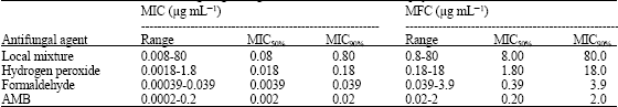
| Fig. 1: | Minimum fungicidal concentration (MFC50 and MFC90) of the antifungal |
 | |
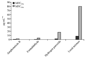
| Fig. 2: | Minimum inhibitory concentration (MIC50% and MIC90%) of antifungal |
Some of these conditions are very different from those recommended by the National Committee for Clinical Laboratory Standards (2002) method for filamentous fungi. In this study, all the antifungal drugs tested, with the exception of the local mixture, displayed excellent activity. We measured MFCs with a simple but rigorous method requiring complete elimination of viable particles in the culture well during the MIC incubation time, while MFC is defined as a = 99% reduction of CFU (Espinel-Ingroff, 2001; Espinel-Ingroff et al., 2002; Moore et al., 1993; Pujol et al., 2000; Ali-Shtayeh et al., 1988). Evaluation of antifungal drugs results in Fig. 1 and 2 show that AMB was more effective than other anti-fungal agents with MIC90 at the concentration of 0.02 μg mL-1 and MFC90 was at the concentration of 2.0 μg mL-1, followed in second place by Formaldehyde, where, MIC90 was at the concentration of 0.039 μg mL-1 and it's MFC90 was at the concentration of 3.9 μg mL-1. Followed in third place was hydrogen peroxide solution where the MIC90 was at the concentration of 0.18 μg mL-1 and the MFC90 was at the concentration of 18 μg mL-1. The local mixture was at fourth place, where, the MIC90 was at the concentration of 0.8 μg mL-1 and the MFC90 was at the concentration of 80.0 μg mL-1.
CONCLUSIONS
This study showed that the effective and most efficient drugs to eliminate the fungus T. verrucosum was AMB, followed by 10% formaldehyde and hydrogen peroxide solution, The least effective was the local mixture. This is in agreement with the findings of Espinel-Ingroff (2002) and Wildfeuer et al. (1998) who found that the anti-fungal agent AMB to be effective on skin fungus and fungi un-common. Also, we agree with the conclusions of Reinner (1992) who found that calves infected with ringworm could be treated with intravenous injection of 10% formaldehyde. Present results agree with those of other authors (William et al., 1980), who demonstrated that susceptibility to formaldehyde (5%) and sodium iodide (1%) in combination when were applied topically to successfully treat dermatophytosis in fox. Other researchers evaluated a local mixture in Jordan of the antifungal component of salicylic acid, benzoic acid, sulfur and iodine; Showing positive results of the drug for the topical treatment of this disease (Al-Ani et al., 1995; Soares and Cury, 2001; Allen et al., 1982). The importance of oxidative products of the respiratory burst on the killing of bacterial, parasitic and fungal organisms has been well established (Rosen and Klebanoff, 1979; Dockrell and Playfairj, 1984; Lehrer, 1969; Babior, 1978). In particular, the Klebanoff system (Klebanoff, 1968) comprising the reaction between hydrogen peroxide, peroxidase (myeloperoxidase) and a halide is regarded as one of the major components of anti-microbial host defence mechanisms. However, present study showed that hydrogen peroxide came in third place in term of effectiveness against the dermatophyte. In general different pharmacological treatment have been recommended to control dermatophytes; Terbinafine (TF), Itraconazole (ITZ), Fluconazole (FCZ) and more recently, Voriconazole (VCZ) and the new triazole UR-9825, still under clinical investigation (Aly, 1997; Agwa et al., 2000; Mock et al., 1998; Rex et al., 1997; Gupta et al., 2001; Tawara et al., 2000). These drugs, produce their therapeutic effects by disrupting the structure and function of various fungal cell components . Polyenes (e.g., AMB) and azoles (e.g., fluconazole) act on ergosterol to disrupt fungal cell membranes. Both types of drugs also affect cholesterol in human cell membranes and this characteristic is considered primarily responsible for the drugs’ toxicities. Recently, the use of natural plant products (garlic, lemon grass, datura, acacia, onions, a triplex, ginger, black seed, neem, basil, eucalyptus, alfalfa and basil) has been emerged to localize treatment of T. verrucosum that causes ringworm in calves (Saadabi, 2006; Omar and Abd-El-Halim, 1992; Aly et al., 2000); Aly and Bafiel, 2008; Koneman and Roberts, 1985; Clark et al., 1990; Jain et al., 2004). A study demonstrated the effectiveness of the Lawsonia inermis - Henna plant after drying and milling treatment of localized disease ringworm in calves (Bosoglu et al., 1998). Others (Sarkar, 1986) showed that Euphorbia thymifolia leaf treated the T. verrucosum infected calves. They are safe to human and the ecosystem than the chemical antifungal compounds (Shelef, 1983). Development of more effective and less toxic antifungal agents is required for the treatment of dermatophytosis. In summary the proposed microdilution assay for dermatophytes is convenient and reproducible in contrast to others (Meletiadis et al., 2000). However, it is important to obtain more clinical data to confirm if this in vitro efficacy is predictive for clinical results. Among, the antifungal tested, AMB was the most potent, while the local mixture was the least active topical agent.
ACKNOWLEDGMENT
We acknowledge and thank Abdulatif Alsagar for cooperation and help in the study.
REFERENCES
- Abdullah, S.K. and D.A. Hassan, 1995. Isolation of dermatophytes and other keratinophilic fungi from surface sediments of the Shatt Al-Arab River and its creeks at Basra, Iraq. Mycoses, 38: 163-166.
Direct Link - Ali-Shtayeh, M.S., H.M. Arda, M. Hassouna and S.F. Shaheen, 1988. Keratinophilic fungi on the hair of cows, donkeys, rabbits, cats and dogs from the West Bank of Jordan. Mycopathologia, 104: 109-121.
Direct Link - Araujo, C.R., K.C. Miranda, O.F.L. Fernandes, A.J. Soares and M.R. Silva, 2009. In vitro susceptibility testing of dermatophytes isolated in Goiania, Brazil, against five antifungal agents by broth microdilution method. Rev. Insti. Med. Trop. South Paulo, 51: 9-12.
Direct Link - Abou-Gabal, M., G.A. El-Galil, A. El-Nore and D.A. El-Rehim, 1976. Animal ringworm in Upper Egypt. Sabouraudia, 14: 33-36.
CrossRefDirect Link - Ali-Shtayeh, M.S., 1989. Keratinophilic fungi of schools playgrounds in the nablus area, west bank of Jordan. Mycopathologia, 106: 103-108.
CrossRefDirect Link - Al-Ani, F.K., L.S. Al-Bassam and K.A. Al-Salahi, 1995. Epidemiological study of dermatomycosis due to Trichophyton schoenleinii in camels in Iraq. Bull. Anim. Health Prod. Africa, 43: 87-92.
Direct Link - Ajello, L., 1974. Natural history of the dermatophytes and related fungi. Mycopathologia, 53: 93-110.
CrossRefDirect Link - Allen, H.B., P.J. Honig, J.J. Leyden and K.J. McGinley, 1982. Selenium sulfide: Adjunctive therapy for tinea capitis. Pediatrics, 69: 81-83.
Direct Link - Babior, B.M., 1978. Oxygen-dependent microbial killing by phagocytes. N. Engl. J. Med., 298: 659-668.
CrossRefDirect Link - Barchiesi, F., D. Arzeni, V. Camiletti, O. Simonetti, A. Cellini, A.M. Offidani and G. Scalise, 2001. In vitro activity of posaconazole against clinical isolates of dermatophytes. J. Clin. Microbiol., 39: 4208-4209.
CrossRefDirect Link - Berger, A.R., 1996. Common superficial fungal infections in patient with AIDS. Clin. Infec. Dis., 22: 128-132.
Direct Link - Bosoglu, A., F. Birdane and H. Solmaz, 1998. The effect of henna (Folium) Lawsonia] past in ringworm in valves. Indian Vet. J., 75: 71-72.
Direct Link - Butty, P., J.C. Lebecq, M. Mallie and J.M. Bastide, 1995. Evaluation of the susceptibility of dermatophytes to antifungal drugs: A new technique. J. Med. Vet. Mycol., 33: 403-409.
Direct Link - Chermette, R., L. Ferreiro and J. Guillot, 2008. Dermatophytoses in animals. Mycopathologia, 166: 385-405.
CrossRefDirect Link - Clark, A.M., T.M. Jurgens and C.D. Hufford, 1990. Antimicr-bial activity of juglone. Phytotherpay Res., 4: 11-14.
Direct Link - Del-Rosso, J.Q., 2000. Current management of onychomycosis and dermatomycoses. Curr. Infec. Dis. Rep., 2: 438-445.
Direct Link - Dockrell, H.M. and H.L. Playfairj, 1984. Killing of plasmodium yoelii by enzyme-induced products of the oxidative burst. Infec. Immunol., 43: 451-456.
Direct Link - Espinel-Ingroff, A., 2001. In vitro fungicidal activities of voriconazole, itraconazole, and amphotericin B against opportunistic moniliaceous and dematiaceous fungi. J. Clin. Microbiol., 39: 954-958.
Direct Link - Espinel-Ingroff, A., A. Fothergill, J. Peter, M.G. Rinaldi and T.J. Walsh, 2002. Testing conditions for determination of minimum fungicidal concentrations of new and established antifungal agents for Aspergillus spp.: NCCLS collaborative study. J. Clin. Microbiol., 40: 3204-3208.
Direct Link - FadlElmula, A., H. Agab, J.M. LeHorgene, B. Abbas and A.E. Abdalla, 1994. First isolation of Trichophyton verrucosum as the etiology of ringworm in the sudanese camels (Camelus dromedarius). Rev. Elev. Med. Vet. Pays. Trops., 47: 184-187.
Direct Link - Fernandez-Torres, B., A.J. Carrillo, E. Martin, A. Del Palacio and M.K. Moore et al., 2001. In vitro activities of 10 antifungal drugs against 508 dermatophyte strains. Antimicrob. Agents Chemother., 45: 2524-2528.
Direct Link - Fung-Tome, J.C., B. Minassian, E. Huczko, B. Kolek, D.P. Bonner and R.E. Kessler, 1995. In vitro antifungal and fungicidal spectra of a new pradimicin derivative, BMS –181184. Anti Microbial Agents Chemotherapy, 39: 295-300.
Direct Link - Gehrt, A., J. Peter, P.A. Pizzo and T.J. Walsh, 1995. Effect of increasing inoculum sizes of pathogenic filamentous fungi on MICs of antifungal agents by broth microdilution method. J. Clin. Microbiol., 33: 1302-1307.
Direct Link - Gitao, C.G., H. Agab and A. Khalifalla, 1998. An outbreak of a mixed infection of Dermatophilus congolensis and Microsporum gypseum in camels (Camelus dromedarius) in Saudi Arabia. Rev. Sci. Tech. Off. Int. Epiz., 17: 749-755.
PubMed - Gupta, A.K., P. Adam, N. Dlova, C.W. Lynde and S. Hofstader et al., 2001. Therapeutic options for the treatment of tinea capitis caused by Trichophyton species: Griseofulvin versus the new oral antifungal agents, terbinafine, itraconazole and fluconazole. Pediatr. Dermatol., 18: 433-438.
Direct Link - Hazen, K.C., 1998. Fungicidal versus fungistatic activity of terbinafine and itraconazole: An in vitro comparison. J. Am. Acad. Dermatol., 38: S37-S41.
Direct Link - Hazen, K.C., 2000. Evaluation of in vitro susceptibility of dermatophytes to oral antifungal agents. J. Am.Acad. Dermatol., 43: 125-129.
Direct Link - Hofbauer, B., I. Leitner and N.S. Ryder, 2002. In vitro susceptibility of Microsporum canis and other dermatophyte isolates from veterinary infections during therapy with terbinafine or griseofulvin. Med. Mycol., 40: 179-183.
Direct Link - Jain, N., M. Sharma and P. Kumar, 2004. Regulatory effect of some plant extracts on the growth of dermatophytic fungi. Indian J. Microbiol., 44: 59-62.
Direct Link - Jessup, C.J., J. Warner, N. Isham, I. Hasan and M.A. Ghannoum, 2000. Antifungal susceptibility testing of dermatophytes: Establishing a medium for inducing conidial growth and evaluation of susceptibility of clinical isolates. J. Clin. Microbiol., 38: 341-344.
PubMedDirect Link - Jessup, C.J., M.A. Ghannoum and N.S. Ryder, 2000. An evaluation of the in vitro activity of terbinafine. Med. Mycol., 38: 155-159.
Direct Link - Korting, H.C., M. Ollert and D. Abeck, 1995. Results of german multicenter study of antimicrobial susceptibilities of Trichophyton rubrum and Trichophyton mentagrophytes strains causing tinea unguium. Antimicrob. Agents Chemotherapy, 39: 1206-1208.
Direct Link - Klebanoff, S.J., 1968. Myeloperoxidase-halide-hydrogen peroxide antibacterial system. J. Bacteriol., 95: 2131-2138.
PubMedDirect Link - Koneman, E.W. and G.D. Roberts, 1985. Practical Labratory Mycology. 3rd Edn., wolliams and Wolkins, Baltimore, ISBN-13: 978-0683047462, pp: 216.
Direct Link - Lehrer, R.I., 1969. Antifungal effects of peroxidase systems. J. Bacteriol., 99: 361-365.
Direct Link - Macura, A., 1995. Dermatophytes, pathogens or saprophytes. Int. J. Dermatol., 34: 529-530.
Direct Link - Mahmoudabadi, A.A. and M. Zarrin, 2008. Isolation of dermatophytes and related keratinophilic fungi from the two public parks in Ahvaz. Jundishapur J. Microbiol., 1: 20-23.
Direct Link - Manavathu, E.K., J. Cutright and P.H. Chandrasekar, 1999. Comparative study of susceptibilities of germinated and ungerminated conidia of Aspergillus fumigatus to various antifungal agents. J. Clin. Microbiol., 37: 858-861.
Direct Link - Meletiadis, J., J.F.G.M. Meis, J.W. Mouton, J.P. Donnelly and P.E. Verweij, 2000. Comparison of NCCLS and 3-(4,5-dimethyl-2-thiazyl)-2,5-diphenyl-2H-tetrazolium bromide (MTT) methods of in vitro susceptibility testing of filamentous fungi and development of a new simplified method. J. Clin. Microbiol., 38: 2949-2954.
Direct Link - Mock, M., M. Monod, F. Baudruz-Rosselet and R.G. Panizzon, 1998. Tinea capitis dermatophytes: Susceptibility to antifungal drugs tested in vitro and in vivo. Dermatology, 197: 361-367.
Direct Link - Moore, C.B., D. Law and D.W. Denning, 1993. In-vitro activity of the new triazole D0870 compared with amphotericin B and itraconazole against Aspergillus spp. J. Antimicrobial Chemotherapy, 32: 831-836.
Direct Link - Niewerth, M., V. Splanemann, H.C. Korting, J. Ring and D. Abeck, 1998. Antimicrobial susceptibility testing of dermatophytes—comparison of the agar macrodilution and broth microdilution tests. Chemotherapy, 44: 31-35.
Direct Link - Norris, H.A., B.E. Elewski and M.A. Ghannoum, 1999. Optimal growth conditions for the determination of the antifungal susceptibility of three species of dermatophytes with the use of a microdilution method. J. Am. Acad. Dermatologists, 40: S9-S13.
Direct Link - Nimura, K., Y. Niwano, S. Ishiduka and R. Fukumoto, 2001. Comparison of in vitro antifungal activities of topical antimycotics launched in 1990s in Japan. Int. J. Antimicrobial Agents, 18: 173-178.
Direct Link - Perea, S., A.W. Fothergill, D.A. Sutton and M.G. Rinaldi, 2001. Comparison of in vitro activities of voriconazole and five established antifungal agents against different species of dermatophytes using a broth macrodilution method. J. Clin. Microbiol., 39: 385-388.
Direct Link - Pujol, I., C. Aguilar, J. Fernandez-Ballart and J. Guarro, 2000. Comparison of the minimum fungicidal concentration of amphotericin B determined in filamentous fungi by macrodilution and microdilution methods. Med. Mycol., 38: 23-26.
Direct Link - Rex, J.H., M.A. Pfaller, J.N. Galgiani, M.S. Bartett and A. Espinel-Ingroff et al., 1997. Development of interpretive breakpoints for antifungal susceptibility testing: Conceptual framework and analysis of in vitro-in vivo correlation data for fluconazole, itraconazole and Candida infections. Clin. Infect. Dis., 24: 235-247.
CrossRefDirect Link - Rosen, H. and S.J. Klebanoff, 1979. Bactericidal activity of a superoxide anion-generating system. A model for the polymorphonuclear leukocyte. J. Exp. Med., 149: 27-39.
Direct Link - Saadabi, A.M.A., 2006. Antifungal activity of some saudi plants used in traditional medicine. Asian J. Plant Sci., 5: 907-909.
CrossRefDirect Link - Tawara, S., F. Ikeda, K. Maki, Y. Morishita and K. Otomo et al., 2000. In vitro activities of a new lipopeptide antifungal agent, FK463, gainst a variety of clinically important fungi. Antimicrob. Agents Chemotherapy, 44: 57-62.
Direct Link - Tsang, P., T. Hopkins and V. Jimenez-Lucho, 1996. Deep dermatophytosis caused by Trichophyton rubrum in a patient with AIDS. J. Am.Acad. Dermatology, 34: 1090-1091.
Direct Link - Venugopal, P.V. and T.V. Venogopal, 1992. Superficial mycoses in Saudi Arabia. Aust. J. Dermatol., 33: 45-48.
CrossRefDirect Link - William, U.K., C.E. Gates and G.H. Ruth, 1980. Trichophyton mentagrophytes dermatophytosis in wild fox. J. Wildlife Dis., 16: 465-468.
PubMedDirect Link - Wildfeuer, A., H.P. Seidl, I. Paule and A. Haberreiter, 1998. In vitro evaluation of voriconazole against clinical isolates of yeasts, moulds and dermatophytes in comparison with itraconazole, ketoconazole, amphotericin B and griseofulvin. Mycoses, 41: 309-319.
Direct Link