
Ashraf M. Abu-Seida
Department of Surgery, Anesthesiology and Radiology, Faculty of Veterinary Medicine, Cairo University, P.O. Box 12211, Giza, Egypt
LiveDNA: 20.4481
ORCID: 0000-0001-5466-2016
Asian Journal of Animal Sciences
Year: 2015 | Volume: 9 | Issue: 2 | Page No.: 80-84


ABSTRACT
Cutaneous squamous cell carcinoma is of great economic importance in sheep producing countries. The present report records-for the first time-a case of congenital cutaneous squamous cell carcinoma in a 10-day-old male lamb. The neoplasm appeared as an ulcerated, blackish tumor of bad odor at the left supra-orbital area. The sub-mandibular and pre-scapular lymph nodes were swollen. Corneal opacity and purulent ocular discharge were also observed in the related eye. Cut section of the neoplasm revealed reddish white, fleshy moist surface with ulcerated blackish overlying layer. Microscopically, the neoplasm had numerous keratin pearls, extensive fibrous stroma and leucocytic cell infiltration mainly neutrophils and lymphocytes. The neoplastic cells showed mitotic figures. Surgical excision of the neoplasm under local infiltration analgesia was curative without recurrence. In conclusion, ovine cutaneous squamous cell carcinoma may occur as a congenital affection.
PDF Abstract XML References Citation
Received: February 20, 2015;
Accepted: April 06, 2015;
Published: April 13, 2015
How to cite this article
Ashraf M. Abu-Seida, 2015. Congenital Cutaneous Squamous Cell Carcinoma in a Lamb. Asian Journal of Animal Sciences, 9: 80-84.
DOI: 10.3923/ajas.2015.80.84
URL: https://scialert.net/abstract/?doi=ajas.2015.80.84
DOI: 10.3923/ajas.2015.80.84
URL: https://scialert.net/abstract/?doi=ajas.2015.80.84
INTRODUCTION
Sheep constitute an important part of the livestock in Egypt, producing, meat, milk, hide and wool (Zabady et al., 2004). Skin lumps were commonly reported in sheep causing damage to the hide and wool (Abu-Seida, 2014).
Cutaneous squamous cell carcinoma is a malignant tumor of epithelial origin, deriving from keratinocytes that widely reported in domestic animals (Yager and Scott, 1993; Zabady et al., 2004; Abu-Seida and Kawkab, 2007).
This neoplasm was prevalent in adult and old sheep especially white coated animals (Mendez et al., 1997; Zabady et al., 2004). Eyelids, vulva and perineal region were the most commonly affected regions (Vandegraaff, 1976; Steven and Stoops, 1988).
Economically, this neoplasm was listed as a major cause of losses in adult sheep (Bush et al., 2006) especially in summer and autumn (Mendez et al., 1997).
Multifactorial etiology was suggested for development of this neoplasm including; poor skin pigmentation and prolonged exposure to ultra-violet radiation (Vandegraaff, 1976; Steven and Stoops, 1988).
Clinically, the initial lesions were non-specific and consisted of erythema, hyperkeratosis, actinic keratosis and dermatitis. The tumors grew slowly but progressively over a period of 1-2 years, with frequent complications by bacterial infections or secondary myiasis (Mendez et al., 1997).
Histopathological examination of the tumor revealed well-differentiated squamous cell carcinomas, surrounded by a moderate to abundant infiltrate of lymphocytes, plasma cells and macrophages. The overlying epidermis was frequently ulcerated and inflamed (Mendez et al., 1997).
Although several congenital affections were recorded in sheep, cutaneous squamous cell carcinoma has never been reported before (Senna et al., 2003; Senna and Abu-Seida, 2004). Therefore, the aim of the present report was to put in records-for the first time-a rare case of congenital cutaneous squamous cell carcinoma in a lamb.
MATERIALS AND METHODS
History: A ten-day-old male lamb was admitted to the surgery clinic at Faculty of Veterinary Medicine, Cairo University, Egypt for treatment of supra-orbital overgrowth.
Case history revealed that the lamb had born with a congenital supra-orbital outgrowth which grew progressively.
Surgical excision: The neoplasm was excised surgically under local infiltration analgesia using 3 mL of lidocaine HCl 2% solution (Lidocaine®, Hospira Co., Lake Forest, IL). Hemostasis was done by tampon then the skin was closed by silk #2/0 using simple interrupted pattern. The affected eye was treated by local ophthalmic Tobramycin and Dexamethasone drops (Tobradex® eye drops, EIPICO, Egypt) for 1 week.
The size and weight of the excised neoplasm were measured and multiple specimens were fixed in 10% formalin solution, sectioned at 4-5 μm thickness and stained with hematoxylin and eosin (Bancroft and Gamble, 2013).
RESULTS
Clinical signs: Clinical examination revealed hard, ulcerated, blackish tumor of bad odor at the left supra-orbital area (Fig. 1). The sub-mandibular and pre-scapular lymph nodes were swollen. The lamb had a white coat while the head was black. Corneal opacity and purulent ocular discharge were also observed.
Histopathological findings: Macroscopically, the size of excised neoplasm was 3×2 cm (Fig. 2a) and weight was 30 g. Cut section of the neoplasm revealed reddish white, fleshy moist surface with ulcerated blackish overlying layer (Fig. 2b).
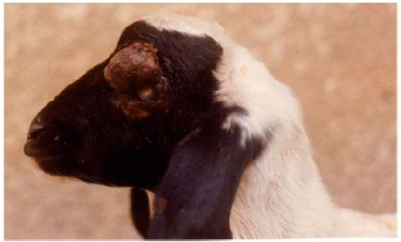 | |
| Fig. 1: | A ten-day-old lamb showing left supra-orbital cutaneous squamous cell carcinoma. Notice the left corneal opacity |
 | |
| Fig. 2(a-b): | (a) Excised neoplasm showing blackish ulcerated surface. (b) Cut section of the excised neoplasm showing reddish white moist surface |
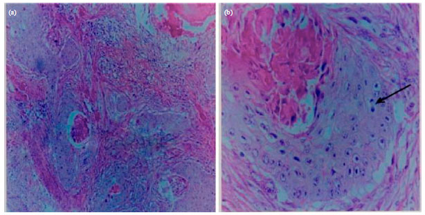 | |
| Fig. 3(a-b): | (a) Photomicrograph of the excised neoplasm showing keratin pearls, extensive fibrous stroma and leucocytic cells infiltration (H and Ex100). (b) Photomicrograph of the excised neoplasm showing neoplastic epithelial cells with mitotic figures (arrow) (H and Ex400) |
Microscopically, the neoplasm had numerous keratin pearls, extensive fibrous stroma and leucocytic cell infiltration mainly neutrophils and lymphocytes (Fig. 3a). The neoplastic cells showed mitotic figures (Fig. 3b). The skin showed ulcerative dermatitis.
Surgical excision of the neoplasm was curative without recurrence. The corneal opacity was subsided within 2 weeks.
DISCUSSION
Cutaneous squamous cell carcinoma is of great economic importance in sheep producing countries as it affects the produced wool and hide grade.
Reports concerning this neoplasm in sheep are sparse with little documentation of its etiology, prevalence and sites. In addition, there is no report documented this neoplasm as a congenital affection. Therefore, this report documents for the first time a case of congenital cutaneous squamous cell carcinoma in a lamb.
Regarding the age, it is well known that cutaneous squamous cell carcinoma usually affects adult sheep especially ewes over 5 years old (Mendez et al., 1997; Zabady et al., 2004). In contrast, the age of affected male lamb was 10 days.
Cutaneous squamous cell carcinoma usually affects white coated sheep (Steven and Stoops, 1988; Mendez et al., 1997). Although, the reported lamb had white coat, the affected area was black.
The most common sites of cutaneous squamous cell carcinoma in sheep were; eyelids, vulva, perineum and stifle fold (Steven and Stoops, 1988; Mendez et al., 1997; Fouad et al., 2000; Zabady et al., 2004). The aforementioned regions have no or little wool and continuously exposed to sunlight therefore, excessive exposure to sunlight, lack of melanin pigment and razing in arid or semiarid regions have been incriminated as predisposing causes for cutaneous squamous cell carcinoma in sheep. This report added the supra-orbital area to the aforementioned affected regions. The occurrence of the recorded neoplasm at birth and in a black area means that this incrimination may be incorrect.
The bad odor of the neoplasm could be attributed to secondary bacterial infections. Mendez et al. (1997) added dermal myiasis as a complication of long standing cases.
Regional lymph nodes of the affected lamb were enlarged as a result of secondary bacterial infection but not because of metastasis. A finding that was confirmed by histopathological section which revealed the presence of inflammatory cells mainly neutrophils. Nuttall et al. (1990) mentioned that regional lymph nodes had no influence on the growth of ovine squamous cell carcinoma. However, Steven and Stoops (1988) found metastatic lesion to the regional lymph node and lungs.
In addition, the corneal opacity and purulent ocular discharge of the related eye could be attributed to the severe keratitis induced by pressure of neoplasm and extension of secondary bacterial infection to the affected eye.
The characteristic pathological findings of cutaneous squamous cell carcinoma included keratin pearls (cell nest), large neoplastic cells with large nucleus and mitotic figures. Similar findings were mentioned by Steven and Stoops (1988) and Mendez et al. (1997).
Surgical excision of the neoplasm was curative without recurrence. Similar findings were mentioned by Fouad et al. (2000).
In conclusion, ovine cutaneous squamous cell carcinoma may be occurring as a congenital affection.
ACKNOWLEDGMENT
The author would like to acknowledge Prof. Dr. Kawkab A. Ahmed, professor of pathology at Faculty of Veterinary Medicine, Cairo University for her grateful help in the histopathological examination.
REFERENCES
- Abu-Seida, A.M., 2014. Radiographical examination and treatment of wattle cyst in goats and sheep. Global Vet., 12: 862-864.
Direct Link - Bush, R.D., J.A. Toribio and P.A. Windsor, 2006. The impact of malnutrition and other causes of losses of adult sheep in 12 flocks during drought. Aust. Vet. J., 84: 254-260.
CrossRef - Nuttall, W.O., P.W. Daniels and P.W. Ladds, 1990. Non-effect of regional lymph node removal on growth of ovine aural squamous cell carcinoma. J. Comp. Pathol., 103: 215-219.
CrossRef - Senna, N.A., A.M.A. Abu-Seida, S.M. Gadallah, Inas, N. El-Husseiny and G.M. Rakha, 2003. Congenital anomalies in native breeds of sheep and goats: A report on 120 cases of 24 varieties. Vet. Med. J. Giza., 51: 363-380.
Direct Link - Vandegraaff, R., 1976. Squamous-cell carcinoma of the vulva in merino sheep. Aust. Vet. J., 52: 21-23.
CrossRef