
Nael M. Fawzi
Department of Biology, Faculty of Science, UAE University, Al Ain, UAE
Research Journal of Botany
Year: 2011 | Volume: 6 | Issue: 2 | Page No.: 68-77


ABSTRACT
Seed characteristics of 12 species representing 7 genera of Caesalpinioideae, were studied in order to define the diversity in the characters of the shape, areole, micropyle, hilum and lens as well as the testa sculpturing patterns by using both a light and Scanning Electron Microscope (SEM). The results of SEM investigation of seed coat sculpturing exhibited five distinct types of surface patterns namely; levigate, substriate, reticulate, rugulate and polygonal-discoid. Valuable taxonomic evidence has been obtained from studying the seed characteristics. Many of these characteristics are diagnostic at both the generic and specific levels.
PDF Abstract XML References Citation
Received: February 28, 2011;
Accepted: April 14, 2011;
Published: June 23, 2011
How to cite this article
Nael M. Fawzi, 2011. Macro-and Micromorphological Seed Characteristics of some Selected Species of Caesalpinioideae-Leguminosae. Research Journal of Botany, 6: 68-77.
URL: https://scialert.net/abstract/?doi=rjb.2011.68.77
URL: https://scialert.net/abstract/?doi=rjb.2011.68.77
INTRODUCTION
Leguminosae comprises three subfamilies namely; Caesalpinioideae, Mimosoideae and Papilionoideae (Rendle, 1952; Willis, 1976). However, Hutchinson (1948) promoted the rank of Leguminosae to that of the order “Leguminales”, which in turn comprises the three families: Caesalpiniaceae, Mimosaceae and Fabaceae (Papilionaceae). The subfamily Caesalpinioideae includes 180 genera and 3000 species of tropical and subtropical trees and shrubs (Heywood, 1971). The largest genus is Cassia with about 600 species chiefly centered in the Pantropical region (Brenan, 1967).
Seed coat characters are successfully employed in the identification and classification of taxa by Erol et al. (2006), Kaplan et al. (2007), Jafari et al. (2009) and Fawzi et al. (2010). Taia (2004) referred that the SEM helps to detect minute taxonomically significant structures in seed coat patterns which might enable us to define species characteristics. Seed morphology and structure in the Leguminosae has been the subject of numerous studies by Corner (1951, 1976), Gunn (1972, 1981a, b), Hanna (1984), Manning and Van Staden (1985), Lersten et al. (1992), Jones and Geneve (1995), Lu-Ayaz et al. (2000), Hussein et al. (2002a, b) and Salimpour et al. (2007).
Manning and Van staden (1987) showed that the cuticle sculpturing in Caesalpinioideae is rather conservative in diversity and is usually rugose, rarely with rugae overlain with a reticulum. They suggested that the expression of cuticular sculpturing is related to the proximity of the underlying epidermal cells, the influence of which is attenuated when exerted over distances greater than 10 μm. They recorded the occurrence of fracture lines (cracks) in all taxa examined. Other studies have reported a somewhat greater diversity (Bragg and Bridges, 1984).
Gunn (1991) provided a morphological description of seeds including lens (which is a part of seed coat but modified) data for 148 species in the Caesalpinioideae. He reported that lens shapes were categorized as circular, elliptic, irregular, linear, oblong, punctiform, square, triangular or wedge shaped; the lens may be raised (a mound), flush or recessed. Hamly (1932), Aitken (1939) and Hagon and Ballard (1970) reviewed the structure of the legume seeds with emphasis to the role of the lens. They recorded that hard seeds of legumes become permeable through the propensity of the lens palisade to split under pressure. Staden et al. (1989) reported that the lens is the limiting inbuilt point of weakness in absolutely hard seeds but the lens and testa should be regarded as an integrated system.
Sahai (1999) investigated the details of structural diversity of lens in 14 species of Cassia in relation to taxonomic significance. She recorded that each species is characterized by its own lens. Moreover, shape, size and position of the lens in each species are genetically constant and serve as distinction from allied species of same genus. Sahai et al. (1997) carried out the macro- and micromorphology of seeds of 21 Cassia species and showed large variation in seed morphology.
This study focused the light upon the diversity in the characteristics of the shape, areole, micropyle, hilum and lens as well as the testa surface patterns of seeds in twelve species belonging 7 genera of the Caesalpinioideae.
MATERIALS AND METHODS
In the present study, twelve taxa of the subfamily Caesalpinioideae have been investigated. Seeds of these species were received, during 2008, from the Royal Botanic Gardens, Century Seed Bank, Kew, England. Species investigated are the following:
| • | Afzelia africana Smith. ex Pers |
| • | Caesalpinia mollis (Kunth) Sprengel |
| • | Caesalpinia pyramidalis Tul |
| • | Chamaecrista nigricans (Vahl) Greene |
| • | Erythrophleum africanum (Welw. ex Benth.) Harms |
| • | Hoffmannseggia lactea (Schinz) Schinz |
| • | Piliostigma reticulatum (DC.) Hochst |
| • | Senna alata (L.) Roxb |
| • | Senna artemisioides (Gaudich. ex DC.) Randell subsp. zygophylla (Benth.) Randell |
| • | Senna covesii (A. Gray) H. S. Irwin and Barneby |
| • | Senna hirsuta (L.) H.S. Irwin and Barneby |
| • | Senna hirsuta (L.) H.S. Irwin and Barneby var. glaberrima (M.E. Jones) H.S. Irwin and Barneby |
The seed dimensions were measured by Nikon Stereoscopic microscope model SMZ 800 using image analysis software NIS-ELEMENTS D. In addition, the general exomorphological features of the seeds were examined using the same microscope.
The finer morphological details were examined using the Scanning Electron Microscope (SEM) Model JEOL JSM-5600 at the Electron Microscope Unit, United Arab Emirates University. The SEM-micrographs were taken after the mature seeds were coated with a thin layer of gold in JEOL JFC-1200 Fine Coater and examined in different positions using different magnifications.
In case of large seeds which were out of SEM-field viz; Afzelia africana, Caesalpinia mollis, Caesalpinia pyramidalis, Erythrophleum africanum, Piliostigma reticulatum and Senna alata, the whole seeds were photographed using the Stereoscopic microscope.
RESULTS AND DISCUSSION
The shape of seed is usually regular and symmetrical for Mimiosoidaceous and Caesalpinioidaceous seeds. Occasionally, an irregularly shaped seed is found that may be the result of pressure from adjacent seeds (Gunn, 1981b).
From all examined seeds, the seeds of Afzelia africana appeared with the funicle develops into an aril which has a dispersal function and the abscission zone is consequently formed near the base of the funicle leaving the latter attached to the seed during dispersal (Fig. 1). Abscission of seeds normally occurs between the funiculus and the seeds, i.e., the hilum. According to Corner (1976) the term aril is used for a fleshy to hard structure that develops from the funiculus or ovule after fertilization and invests part or all of a seed. The variation in shape of seeds (Table 1), among the species studied, may exhibit a useful clue for identification of the major bulk of the species examined.
Cracks (fracture lines), as in Manning and Van Staden (1987) were recorded in seed surface of all examined taxa at law magnification (less than 30 X). Fracture lines occur in the testa of all caesalpinioid and mimosoid seeds and are apparently functional in seed dehydration (Trivedi et al., 1979).
| Table 1: | The exomorphological characteristic of seeds of the taxa studied of Caesalpinioideae; as viewed under light microscope |
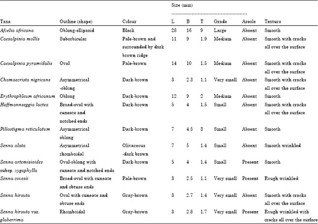 | |
| Grade concerning the size of the seeds. Very small seeds: Less than 5 mm long, Small seeds: 5-10 mm long, Medium sized-seeds: 10-15 mm long, Large seeds: More than 15 mm long. B: Broad, L: Length, T: Thickness | |
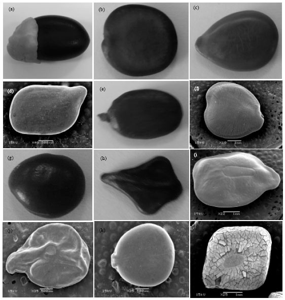 | |
| Fig. 1: | SEM photographs of seed shape: (a) Afzelia africana (b) Caesalpinia mollis (c) Caesalpinia pyramidalis (d) Chamaecrista nigricans (e) Erythrophleum africanum (f) Hoffmannseggia lactea (g) Piliostigma reticulatum (h) Senna alata (i) Senna artemisioides subsp. zygophylla (j) Senna covesii (k) Senna hirsuta and (l) Senna hirsuta var. glaberrima |
According to Gunn (1981a), the Legume testa is usually monochrome brown to black, rarely red, cream or white, or occasionally dichrome as mottling or two distinct coloured areas. In this study, the colour of seeds may, somewhat, be reliable attribute for the recognition of some taxa studied (Table 1). This agrees with the results reported by Shyam and Vartak (1985). They used the colour of seeds with other features to describe the seeds of genus Cassia and to produce a key for their identification. Conversely, Husain (2000) regarded this character to be of limited taxonomic value for its possible fluctuation within the same taxon at the different durations.
Seed size varies from the 3-28 mm long among the studied species (Table 1). The size of seed seems to be diagnostic for Afzelia africana designated, here, as large-sized while the seeds of Senna species are very small sized except S. alata and S. artemisioides subsp. zygophylla are small sized. Consequently, the size of seed may be useful for delimitation at the generic level and also sometimes at the species level. This agrees with the results reported by Hussein et al. (2002a) they adopted the size of seed in some delimitation in Ceasalpinioideae. On the other hand, Thompson (1981) illustrated that the seed size regarded to be of minor taxonomic value and such attribute is subjected to ecological and physiological variations.
The studied seeds of Afzelia, Caesalpinia, Chamaecrista, Erythrophleum, Hoffmannseggia and Piliostigma are without areole. Whereas in Senna, the areole is present in three species, S. hirsuta var. glaberrima, S. covesii and S. artemisioides sub sp. zygophylla (Fig. 1). In this respect, Polhill and Raven (1981) recorded that the hard seeds of Mimosoideae generally have an area on each face (the areole) bounded by a crack in the testa (the pleurogram) which may serve as a valve in drying out the seed and showed that areoles occur sporadically in the Caesalpinioideae. Hussein et al. (2002a) reported that the presence of areole in some species of Senna may be diagnostic while the variation in the areole shape seems to be of very limited taxonomic value.
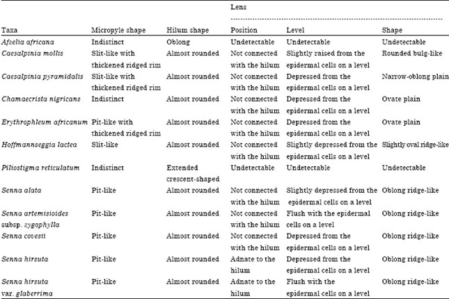
The seeds in the species studied of Caesalpinia, Chamaecrista, Erythrophleum, Hoffmannseggia and Senna are invariably with three apertures; the lens, hilum and micropyle. However, the species studied of Afzelia and Piliostigma, the micropyle and lens are consistently indistinct. These apertures are invariably located at one end of the seed (Fig. 2). Polhill et al. (1981) recorded that the hard seeds in members of Leguminosae invariably have these three apertures. Herein, the lens is consistently found to be located behind the hilum which separates it from the micropyle. The variability in lens characteristics including both the shape and level seems to be quite distinctive for the major bulk of the species investigated (Table 2). It also, presents an additional tool for delimitation of the species examined.
| Table 2: | Characteristic of seeds of taxa studied of Caesalpinioideae; as viewed under scanning electron microscope |
 | |
Furthermore, the lens characteristics shed more light upon the close relationship between the studied species of Senna.
The seeds in the species studied of genus Senna characterized by the presence of both hilum and micropyle in a more or less rounded characteristics plate (Fig. 2). Additionally, the shape of micropyle appears to be unique to Senna species. These results provide additional support for the formerly presumed close relationship between Senna species. Seeds of Piliostigma reticulatum retain hilum shape, crescent-shaped, characterizes this species from the remainder of the taxa examined (Fig. 2). Polhill et al. (1981) referred that in Cercideae-Bauhiniinae, to which the genus Piliostigma was included, a parenchymal connection from the funicle into the seed is apparent as a crescent-shaped line.
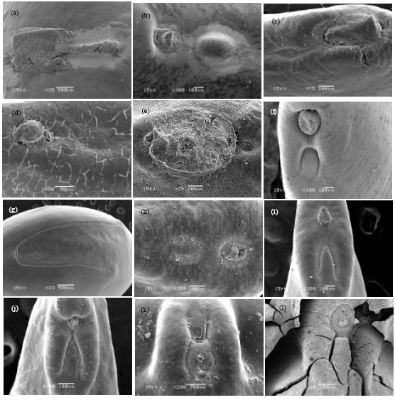 | |
| Fig. 2: | The lens, hilum and micropyle characteristics of seeds among the species studied: (a) Afzelia africana (b) Caesalpinia mollis (c) Caesalpinia pyramidalis (d) Chamaecrista nigricans (e) Erythrophleum africanum (f) Hoffmannseggia lactea (g) Piliostigma reticulatum (h) Senna alata (i) Senna artemisioides subsp. zygophylla (j) Senna covesii (k) Senna hirsuta and (l) Senna hirsuta var. glaberrima |
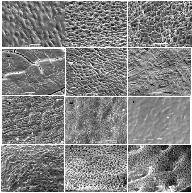 | |
| Fig. 3: | Different patterns of the surface scan of the seeds: (a) Afzelia africana (b) Caesalpinia mollis (c) Caesalpinia pyramidalis (d) Chamaecrista nigricans (e) Erythrophleum africanum (f) Hoffmannseggia lactea (g) Piliostigma reticulatum (h) Senna alata (i) Senna artemisioides subsp. zygophylla (j) Senna covesii (k) Senna hirsuta and (l) Senna hirsuta var. glaberrima |
In this connection, Jones and Genave (1995) showed that such parenchymal connection may serve as a valve to allow water to escape from the seed during desiccation.
The seed surface as viewed under SEM showed five basic sculpturing patterns (Fig. 3) namely; levigate (i.e., smooth), substriate (i.e., irregular striate), rugulate, reticulate and polygonal-discoid. Herein all terminology used for the description of the testa sculpturing patterns are that of Lersten (1981).
Levigate cuticle occurred in Chamaecrista nigricans and Senna artemisioides. But, Chamaecrista nigricans characterized by the presence of many fracture lines and irregular cuticularl folding. Substriate pattern recorded only in Piliostigma reticulatum.
Rugulate pattern with intergraded forms occurred in Erythrophleum africanum (irregularly rugulate with thick rugae), Hoffmannseggia lactea (faintly irregular rugulate), Senna alata (compactly multi-rugulate with fine rugae), Senna covesii (irregular rugulate), Senna hirsuta (rugulate with thick rugae) and Senna hirsuta var. glaberrima (rugulate with spreading irregular depressions).
Reticulate pattern occurred in Caesalpinia mollis (reticulate-foveolate with ridges of the reticulae very thick) and Caesalpinia pyramidalis (irregularly reticulate).
Polygonal-discoid pattern recorded only in Afzelia africana with scattered cuticular deposits.
Herein, the variability in seed surface patterns is seemingly very useful in the recognition of all species studied. This agrees with the results reported by Hussein et al. (2002b). They showed that the finer details of the seed surface appear useful for delimitation at generic level and also species level among the species investigated of Caeasalpinioidea.
In conclusion, it could be declared that valuable taxonomic evidence has been obtained from studying seed characteristics in some species of Caesalpinioidea. Many of these characteristics are diagnostic at both the generic and specific levels.
REFERENCES
- Erol, O., E. Uzen and O. Kucuker, 2006. Preliminary SEM observations on the seed testa structure of Gladiolus L. species from Turkey. Int. J. Bot., 2: 125-127.
CrossRefDirect Link - Fawzi, N.M., A.M. Fawzy and A.A.H.A. Mohamed, 2010. Seed morphological studies on some species of Silene L. (Caryophyllaceae). Int. J. Bot., 6: 287-292.
CrossRefDirect Link - Hanna, P.J., 1984. Anatomical features of the seed coat of Acacia kempeana (Mueller) which relate to increased germination rate induced by heat treatment. New Phytol., 96: 23-29.
CrossRef - Jafari, A., Z. Fathi and M. Bemani, 2009. Using morphology and micromorphology characters for identification of Silene L. species in North-East of Iran. Res. J. Environ. Sci., 3: 667-676.
CrossRefDirect Link - Jones, R.O. and R. Geneve, 1995. Seedcoat structure related to germination in eastern redbud (Cercis canadensis L.). J. Am. Soc. Hort. Sci., 120: 123-127.
Direct Link - Kaplan, A., A. Hasanoglu and I.A. Ince, 2007. Morphological, anatomical and palynological properties of some Turkish Veronica L. species (Scrphulariaceae). Int. J. Bot., 3: 23-32.
CrossRefDirect Link - Lu-Ayaz, S.H. and O.B. lu, 2000. Seed anatomy of five Vicia L. (Leguminosae) species. Pak. J. Biol. Sci., 3: 1440-1442.
CrossRefDirect Link - Polhill, R.M. and P.H. Raven, 1981. Advances in Legume Systematics. Royal Botanical Gardens, Kew, London,.
Direct Link - Sahai, K., 1999. Structural diversity in the lens of the seeds of some Cassia L. (Caesalpinioideae) species and its taxonomic significance. Phytomorphology, 49: 203-208.
Direct Link - Salimpour, F., G. Mostafavi and F. Sharifnia, 2007. Micromorphologic study of the seed of the genus Trifolium, section Lotoidea, in Iran. Pak. J. Biol. Sci., 10: 378-382.
CrossRefPubMedDirect Link - Taia, W.K., 2004. Tribe Trifolieae: Evidence from seed characters. Pak. J. Biol. Sci., 7: 1287-1302.
CrossRefDirect Link - Thompson, P.A., 1981. Variations in seed size within populations of Silene dioica Clairv. In relation to habitat. Ann. Bot., 47: 623-634.
Direct Link - Trivedi, B.S., G.D. Bagchi and V. Bajpai, 1979. Scanning electron microscopic studies on the spermoderm of some Mimosoideae (Leguminosae) India. Phytomorphology, 29: 211-218.
Direct Link