
Samira Omar Abu Baker
Department of Zoology, Girl`s College of Science, University of King Abdul Aziz, Jeddah, Saudi Arabia
Pakistan Journal of Biological Sciences
Year: 2009 | Volume: 12 | Issue: 8 | Page No.: 607-615


ABSTRACT
The present study aimed to investigate the toxic impact of gemcitabine on the histological structure of white mice testis and the histological structure of some embryonic organs. The mature male mice were treated with 130 mg kg-1 of gemcitabine intraperitoneally. These treated males were placed after one week with mature normal females for fertilization. Females were dissected after the 14th day of pregnancy to investigate embryos. The histological investigation of testis showed an interruption in spermatogenesis process as evident by distorted spermatocytes, spermatozoa and a reduction in their number. Histological examination of embryonic organs, including liver, kidney, small intestine, spleen, zatrek gland and testis, after two weeks of pregnancy revealed impaired structures. The drug also reduced fertility and survival of embryos. In conclusion, present study suggests that cautions should be taken when gemcitabine is used as an anticancer drug.
PDF Abstract XML References Citation
How to cite this article
Samira Omar Abu Baker, 2009. Gemcitabine Impacts Histological Structure of Mice Testis and Embryonic Organs. Pakistan Journal of Biological Sciences, 12: 607-615.
DOI: 10.3923/pjbs.2009.607.615
URL: https://scialert.net/abstract/?doi=pjbs.2009.607.615
DOI: 10.3923/pjbs.2009.607.615
URL: https://scialert.net/abstract/?doi=pjbs.2009.607.615
INTRODUCTION
The environmental pollution, chemical products, or food nature may be one of reasons resulted in the great increasing in the number of infected people with cancer (Wogan et al., 2004). The current century develops many ways for cancer treatment either by radial, chemical or surgical treatment. Usage of chemical drugs is the most common way in treatment cancer patients. Thus, researchers are more interest to evaluate the safety of the anti-cancer drugs. Although, these drugs provide some chemical control on this disease, most of these drugs have toxic effect on the normal cells. Unfortunately, up till now, there are no ideal drugs that can damage cells of cancer only without being damage the normal ones. Generally, these drugs damage DNA molecule leading to secondary tumors appearance after chemotherapy ceasing (Magnani et al., 1996). The serious effects of anti-cancer drugs are not only restricted to patients but also to medical workers if they exposed to these drugs for long periods (Brumen and Hovath, 1996). In the present study, one of these new anti-cancer drugs gemcitabine (2, 2 difluoro deoxy cytidine; dfdc) was selected. Gemcitabine is one of the anti-metabolite drugs group. This group consists of three divisions; parapholic acid, para-purine and para-pyrimidine. This group is widely used in chemotherapy and it is efficient in leukemia treatment. Gemcitabine is one of para-pyrimidine that characterized by its efficient on inhibiting the vital formation of the nuclides pyrimidine or resembling their normal metabolism, thus they interfere with the DNA synthesis or on its function.
Para-pyrimidine is widely used; in fungal infections, viral infections contained DNA and cancer diseases (Calabresi and Chabner, 2001). Gemcitabine is also commonly used in treatment of tumor diseases. Earlier studies proved that this drug may lead to apoptosis of lymphatic cancer cells because of DNA partitioning (Huang and Plunkett, 1995) or drug incorporates with DNA. The danger level of this drug increases when some doctors use it with some other drugs (as cisplatin and paciltaxel) that used in chemotherapy to elevate the curial efficiency (Montie, 2005; Brewer et al., 2006). Such drugs are now commonly known that they have toxic effects on the different tissues of the body (Hassan et al., 2005; Goodisman et al., 2006). The anticancer and antineoplastic drugs are known to interfere with DNA and its original substances, so they inhibit DNA synthesis and cause irreversible damage to it (Williams et al., 1985). Usually these drugs have unpleasant effects on the normal tissues as the toxic impacts transfer from the contiguous tumor cells to them by cellular diffusion (Cline and Haskell, 1980). The earlier studies proved that the anti-tumor drugs are often teratogenic or carcinogenic. Also, the anti-metabolites as gemcitabine are considered as anti-cancer drug which interfere with the cell cycle specific of the tumor cells and are more efficient during a certain stage. These drugs are also considered to be toxic for cells in certain phase and known as phase specific drugs. Some of these drugs interfere during the cellular perforation, other during any phase of cell cycle except cells in the G phase. The anti-metabolites have similar structures of the natural anti-metabolites and because of this similarity, some drugs can inhibit enzymes responsible for synthesis of the basic cellular components. Other drugs emit cellular toxic products that fuse with DNA and suppress its function (Lehne et al., 1990).
Gemcitabine is characterized by its high activity against tumor as it is used in the cancer treatment of bladder, blood, ovary, breast, pancreas, soft tissues and neck cancer (Raguse et al., 2005). This drug inhibits the bone marrow function resulting in unusual decrease in number of the white blood cells (leucopenia) and blood platelets (thrombocytopenia), moreover it causes anemia (Aapro et al., 1998). The drug has several effects on the respiratory system causing the toxic respiration (Barlesi et al., 2004), while the kidney failure in patients treated by this drug may be caused due to hemolytic ureic syndrome (Krych, 2005). The cellular toxic effect of gemcitabine is related to its incorporation with cellular DNA molecule, thus it is efficient in inhibition of DNA and RNA synthesis during the cellular perforation phase. This process is known as masked termination where corrected enzymes unable to remove the drug from the binding position (Jordheim et al., 2005). Szatmari et al. (2008) mentioned that the gene expression DCK could prove the toxic impact of gemcitabine in vivo and in vitro when used as an anti-cancer drug. Al-Yamani (2008) concluded that gemcitabine has cytotoxic, clastogenic and mutagenic effect.
Therefore, for the using of gemcitabine in chemotherapy of cancer in USA generally and in KSA particularly (Stucky-Marshall, 1999), as well as the few studies concerned with the toxic effect of this drug, the present study aimed to investigate the impact of gemcitabine on the histological structure of mice testis and embryonic organs.
MATERIALS AND METHODS
Materials: Fifteen of mature male albino mice (age 3 months) were treated by a dose 130 mg kg-1 of gemcitabine through peritoneal injection at August 2008. Then some males were dissected after one week from injection to investigate testis, some others were placed with mature females for fertilization (ratio 1 male:2 female). Females were dissected after the 14th of pregnancy to investigate embryos.
Methods: Treated males were dissected, their testes were removed and specimens of testis were taken for investigation. Pregnant females were dissected to elicitate embryos of two weeks age from pregnancy. Histological sections of different organs of embryos were performed and the percentage of fertility was calculated by pregnant females, survived and died embryos. Specimens from testis and embryonic organs were fixed in 10% of natural formalin and the standard procedures of hydration, clearing and wax embedding were followed. Sections were taken at thickness of 3-5 μm and stained by hematoxylin and eosin (Drury and Wallington, 1980).
RESULTS
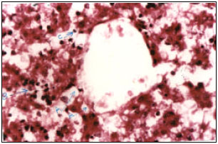
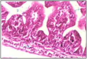
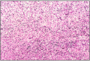
Histological investigation of testis from mature mice treated by gemcitabine: It was observed that fibrosis and analysis the intertubular connective tissue and the number of seminiferous tubules was reduced. The basement membrane of many tubules was appeared decaying or irregularly shape. Some tubules appeared with wide lumen, others expanded and some tubules appeared entirely without spermatocytes and filled with vacuoles. Most tubules were appeared with distorted and necrotic spermatocytes, reduction in the numbers of spermatogonia, secondary spermatocytes and spermatids. While the primary spermatocytes were appeared more densely, distorted, irregular in shape and without nuclei in many of them.
It was also observed that the spermatocytes were not arranged in rows and appeared irregular in shape where they appeared oval, flat or polygonal and rarely rounded. Their cytoplasm was granulated and contains remains of its live components. Most spermatids were observed decayed and others were distorted. Also, the spermatozoa appeared distorted as head or tail disappearance, enlarged head, head separation from tail, short tail, more than one head or changing the head to flat, spindle or pointed shape. However, few numbers of spermatozoa appeared normal, therefore the lumen of most tubules appeared empty. It was also observed that there was not cellular division inside spermatozoa, Ledige cells were found scattered and distorted where they appeared as scattered nuclei and limited presence of sertoli cells with distorted structure (Fig. 1, 2).
Histological investigation of some embryonic organs after two weeks of pregnancy
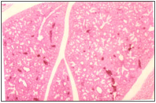
Liver: It was noticed that sections darkly stained with eosin, liver architecture distorted and cellular spatial atrophy of hepatic tissue represented by decayed cellular membrane for most of hepatocytes with scattered and distorted nuclei. Fatty vacuoles can be seen inside and outside the hepatic cells (fatty infiltration) and dilatation of all hepatic acini were observed filled with blood fluid with wide lumens and propagation and thickening of the lining epithelium. Although, the liver did not entirely complete its structure at this age where the hepatic cells were still smaller in size, fewer in number, did not arrange in strands and the portal spaces did not form.
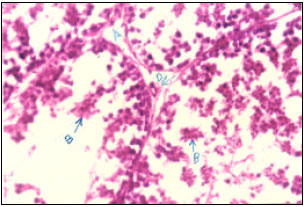 | |
| Fig. 1: | Light micrographs of testis from mature mice treated with gemcitabine showing (A) rare intertubular connective tissue and reduction in number and necrosis of spermatocytes (B) and distortion of Ledig cells (H and E 40x) |
 | |
| Fig. 2: | Light micrographs of testis from mature mice treated with gemcitabine showing (A) distorted primary, (B) secondary spermatocytes, (C) spermatids, (D) spermatozoa and (E) Sertoli cells (H and E 100x) |
It was also observed karyolic nuclei of some hepatocytes, appearance of cytoplasmic rupture with irregular and incomplete cellular membrane. The hepatic cell rarely appeared with normal structure and oedema was observed in the hepatic tissue which represented by liquid infiltration and stasis of blood cells in all blood vessels. Several changes were also recorded as the propagation of the lining epithelium of portal vessels, presence of inflammatory and Kupffer cells, dense lymphatic infiltration and obvious fibrosis of hepatic tissue (Fig. 3-5).
Lung: It was recorded obvious rupture and oedema of lung tissue with disorder in the trabecula walls where the lining epithelium ruptured resulted in their widening in some regions and tightens cavities in most regions of the section.
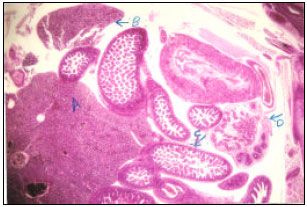 | |
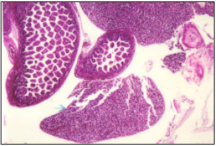
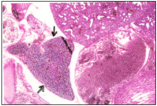
| Fig. 3: | Light micrographs of transverse section of 14 days mice embryo from normal female and male treated with gemcitabine showing (A) liver, (B) spleen, (C) small intestine and (D) testis and epididymus (H and E 10x) |
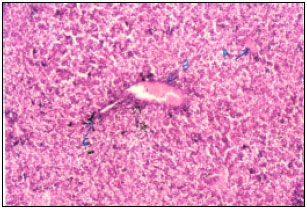 | |
| Fig. 4: | Light micrographs of liver section of 14 days mice embryo from normal female and male treated with gemcitabine showing oedema in the (A) tissue, (B) propagation of the lining epithelium of blood vessels and (C) lymphatic filtration (H and E 40x) |
Vacuoles were spread all over the connective tissue. Regarding the respiratory tracheols, there were rupture in the lining cubic cells with enlargement in their size and propagation in their number led to disconnection with trabecular canals. There was apparent decaying in the columnar lining epithelium of the mucus membrane of the terminal tracheols. It was also observed that folding were fewer in numbers, lower in height, with wider lumen. The muscular layer appeared loosen with obvious vacuoles and the mucus layer appeared with less connective tissue. Obvious rupture of pseudo columnar epithelium, less connective tissue, incomplete hyaline cartilage and submucous layer were observed in the section of trachea (Fig. 6, 7).
 | |
| Fig. 5: | Light micrographs of liver section of 14 days mice embryo from normal female and male treated with gemcitabine showing (A) fatty filtration, (B) lymphatic filtration and (C) Kupffer cells (H and E 100x) |
 | |
| Fig. 6: | Light micrographs of lung section of 14 days mice embryo from normal female and male treated with gemcitabine showing oedema and obvious rupture of the lung tissue (H and E 40x) |
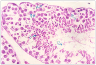
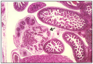
Small intestine: Histological investigation showed disordered of the columnar lining epithelium of the villi which appeared shortly contracted with absence of their cilia and appearing of their basal nuclei in different levels or even inside the villi. Also villi were observed with decayed cellular membranes and walls of most cells leading to fusion and scattering of their nuclei, as well as a reduction in the goblet cells. Crypts of lieberkühn were appeared as cavities surrounded by scattered nuclei where the walls of the lining epithelium were decayed and fused with nuclei of villi scattered cells. Several, histological changes were observed such as oedema in the tissue, stasis of the blood vessels, rarely and presence of connective tissue, decaying of the longitudinal and circular muscular layers and obvious rupture and vacuoles between their fibers (Fig. 8, 9).
 | |
| Fig. 7: | Light micrographs of lung section of 14 days mice embryo from normal (A) female and male treated with gemcitabine showing many vacuoles in the tissue, (B) decaying of the lining epithelium of alveoli, (C) distortion of tracheoles and (D) trachea (H and E 100x) |
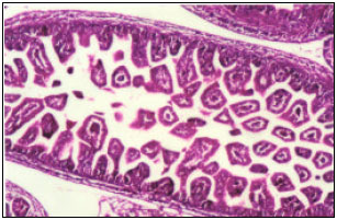 | |
| Fig. 8: | Light micrographs of small intestine section of 14 days mice embryo from normal female and male treated with gemcitabine showing distortion in the histological structure of intestine (H and E 40x) |
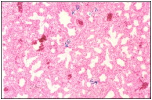
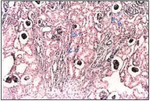
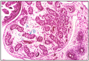
Kidney: Most glomerulus appeared atrophied with rupture of the lining and visceral epithelium and decaying of the blood capillaries. There was also acute decaying in the renal tissue particularly in cortex represented by lessening of the columnar lining cells of the distal tubules, decaying of their cellular walls, scattering their nuclei, enlarging and lessening of the cubic lining cells of the proximal tubules. Moreover, oedema and congestion in the tissue and fibrosis of the connective tissue were also observed. The collecting tubules were also damaged where the lining cells appeared slaughter and the cellular walls of some of them were decayed.
 | |
| Fig. 9: | Light micrographs of small intestine section of 14 days mice embryo from normal female and male treated with gemcitabine showing (A) decaying of the lining epithelium of villi, (B) reducing in goblet cells number and (C) decaying of muscular layer (H and E 100x) |
 | |
| Fig. 10: | Light micrographs of kidney section of 14 days mice embryo from normal female and male treated with gemcitabine showing complete decaying of the renal tissue particularly cortex region (H and E 40x) |
Decaying of the kidney components in some areas led to appearance of wide vacuoles in the tissue (Fig. 10, 11).
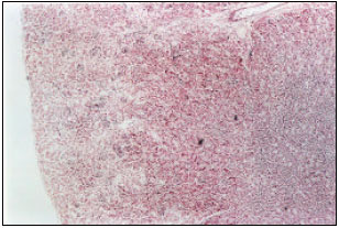
Spleen: Spleen appeared surrounded by connective tissue filled with little number of masses of the lymphatic cells spread on the irregular reticular connective tissue in the splenic strands. Also spleen appeared undivided by barriers of the connective tissue and there were not splenic nodules. Therefore, the white and red core did not form completely in this age. Nearly homogenous coexistence of red blood cells between the lymphatic cells was observed. The size of spleen was clearly larger than that of the control group with oedema and congestion of the tissue, less connective tissue and presence of vacuoles in the tissue (Fig. 12, 13).
 | |
| Fig. 11: | Light micrographs of kidney section of 14 days mice embryo from normal female and male treated with gemcitabine showing (A) glomerulus atrophy, (B) rupture of the lining epithelium of proximal, (C) distal and (D) collecting tubules (H and E 100x) |
 | |
| Fig. 12: | Light micrographs of spleen section of 14 days mice embryo from normal female and male treated with gemcitabine showing oedema in the tissue (H and E 10x) |
Thymes gland: The gland appeared triangular in shape surrounded by fibrous tissue from which barriers expanded inside the gland dividing it into three lobes. The gland lied over the heart composed of dense mass of lymphatic cells among which the red blood cells spread. At this age, there was no cortex or core and the oedema restricted to the blood vessels only. The size of the gland was greater than that of the control group (Fig. 14).
Testis: Testis was surrounded with thin fibrous capsule comparing to the control group. Little number of seminiferous tubules with reduced intertubular connective tissue was appeared in sections.
 | |
| Fig. 13: | Light micrographs of spleen section of 14 days mice embryo from normal female and male treated with gemcitabine showing enlarged spleen and less dense of lymphatic cells (H and E 100x) |
 | |
| Fig. 14: | Light micrographs of zatrek gland section of 14 days mice embryo from normal female and male treated with gemcitabine showing oedema and enlarged gland (H and E 40x) |
The primary spermatocytes appeared distorted in their structure where their walls decayed; their nuclei scattered and filled the lumen of the tubules. Decayed basement membrane was recorded in most seminiferous tubules. Fibrosis and oedema of the connective tissue between and inside tubules were also observed (Fig. 15, 16).
Number of embryos: Table 1 and 2 showed a reduction in fertility percentage after fertilization of normal females by drug treated males. This reduction is due to a diminishing number of spermatozoa, as shown by the histological investigation of testis. The reduction in the number of survived embryos could be explained by the damage effect of the drug on the embryonic tissues, particularly lung, at the developing stages resulting in death of embryos.
 | |
| Fig. 15: | Light micrographs of testis section of 14 days mice embryo from normal female and male treated with gemcitabine (H and E 10x) |
 | |
| Fig. 16: | Light micrographs of testis section of 14 days mice embryo from normal (A) female and male treated with gemcitabine showing fibrous connective tissue, (B) distorted germinal cells and (D) oedema in the tissue (H and E 40x) |
Moreover, drug caused dominant lethal mutation that considered as a genetic change in gametes resulted in embryos death. Such mutation is usually occurring after exposure to chemicals that have a serious effect on the germinal tissue. Generally, the dominant lethal mutation resulted from chromosomal damage (EPA, 1998). Dominant lethal mutation from chemical exposure could be detected by the elevations in the number of died embryos (James and Smith, 1982). This mutation resulted from the disability of chromosome to separate forming monochromosome that led to embryos death at early stage of growth. While trichromosome formation led to embryos death at later stages, thus chemicals influence the vitality and growth of the resulted offspring (Green et al., 1987; Jha et al., 2007).
| Table 1: | Total numbers of died and survived embryos in control group (normal males and females) |
 | |
| Table 2: | Total numbers of died and survived embryos from normal females fertilized by drug treated males |
 | |
DISCUSSION
The pathological changes that appeared in the testicular tissue after injection of male mice with the drug indicate obstructions in the spermatozoa formation and spermiogenesis (transforming of spermatids to spermatozoa processes). These changes could be contributed to the drug toxic effect on the normal cells (Salmon and Apple, 1979).
Gemcitabine is targeting cells in their multiplication phase or any other phase in the cell cycle and damaging their DNA. The toxic effect of gemcitabine is related to its ability to bind with DNA molecule, thus it is efficient in inhibiting DNA synthesis during multiplication phase. It is well known that spermatozoa formation is a series of direct and indirect cellular divisions resulted in cellular multiplication where each germinal cell divided into four spermatids with DNA multiplication. DNA represents the basic genetic material of cells and carries the genetic code that transferred by spermatozoa (Kareem, 1987). These explained the distortion and reduction in numbers of spermatocytes and spermatozoa.
The drug effects on the different embryonic organs may be contributed that this early pregnancy period, is the main period of embryonic organs formation and is considered to be very sensitive period to any external factor (Kareem, 1987). Moreover, there are many studies proved that anti-tumor drugs often cause distorted mutations in embryos as they have the ability to inhibit responsible enzymes of synthesis the main cellular components or they produce toxic substances that later fused with DNA and inhibit its function (Lehne et al., 1990).
The present results of histological investigation of liver are in accordance with those of Mergental et al. (2005) who found that gemcitabine had toxic effect on the liver of rats as causing necrosis of hepatic cells and dense lymphatic filtration that extend to the parenchymal cells surrounded the distorted portal spaces with dense coexistence of inflammatory cells and damaged bile ductules. On the other hand, Kamer et al. (2003) said that gemcitabine usage is safe under certain conditions and its side effects as weight loss is resulted from diarrhea.
This drug obviously damaged all tracheal structures. Gemcitabine affected on the respiratory system and led to sudden contractions of tracheoles, these results were confirmed by Barlesi et al. (2004). In addition, Mergental et al. (2005) found that over dose of gemcitabine resulted in rats’ death and they contributed this to the acute infection of lung. Contrarily, Takatani et al. (2007) found that a dose of 100 mg kg-1 of gemcitabine was very efficient in treatment of lung cancer without any cellular toxicity. This point of view was confirmed by Gagnadoux et al. (2005), who found that a weekly dose of 4 mg kg-1 of gemcitabine was safe for rats as there were no signs of pathological or histological damages particularly in lung except a reduction in the numbers of red blood cells and blood platelets.
The present study found an acute effect of the drug on the kidney that in agreement with Mergental et al. (2005) and Krych (2005), who confirmed that gemcitabine using resulted in kidney failure because of hemolytic uremic syndrome.
Regarding to small intestine, similar effects of the drug found in the present study, were also recognized by Phan et al. (2001), who mentioned that gemcitabine had several toxic effects on intestine especially its mucus layer.
In an experiment carried by Eudaly et al. (1993), who gave pregnant mice females gemcitabine from the 6th to 15th days of pregnancy and dissected females at the 18th day. They found that this drug increases the spleen and zatrek gland weights of pregnant mothers with a reduction in the embryos weight. Oedema, congestion and dense stasis of red blood cells in most tissues may be contributed to the effect of the drug on the bone marrow resulting in unusual reduction in numbers of blood platelets and white blood cells (Aapro et al., 1998). Therefore, we recommended that careful usage of gemcitabine must be taken in consideration and this supported by Mergental et al. (2005) and Zupancic et al. (2007), who assured performing sufficient tests are essential before patients use this drug. We are not with Gang et al. (2007), who confirmed that gemcitabine is safe, effective and successful with less toxicity in cancer tumor therapy.
REFERENCES
- Barlesi, F., P.C. Villani, C. Gimenez and J.P. Kleisbauer, 2004. Gemcitabine-induced severe pulmonary toxicity. Fundament. Clin. Pharmacol., 18: 85-91.
Direct Link - Brewer, C.A., J.A. Blessing, R.A. Nagourney, D.S. McMeekin, S. Lele and S.L. Zweizig, 2006. Cisplatin plus gemcitabine in previously treated squamous cell carcinoma of the cervix: A phase ll study of gynecologic oncology group. Gynecol. Oncol., 100: 385-388.
CrossRef - Brumen, V. and D. Horvath, 1996. Work environment influence on cytostatics-induced genotoxicity in oncology nurses. Am. J. Ind. Med., 30: 67-71.
CrossRefDirect Link - Eudaly, J.A., J.P. Tizzano, G.L. Higdon and G.C. Todd, 1993. Developmental toxicity of gemcitabine, an antimetabolite oncolytic, administered during gestation to CD-1 mice. Teratology, 48: 365-381.
Direct Link - Gagnadoux, F., A. LePape, T. Urban, J. Montharu and L. Vecellio et al., 2005. Safty of pulmonary administration of gemcitabine in rats. J. Aerosol. Med., 18: 198-206.
CrossRefPubMedDirect Link - Goodisman, J., D. Hagrman, K. Tacka and A. Souid, 2006. Analysis of cytotoxicities of platinum compound. Cancer Chem. Pharmacol., 57: 257-267.
CrossRefPubMedDirect Link - Green, S., K.S. Lavappa, M. Manandhar, C.J. Sheu, E. Whorton and J.A. Springer, 1987. Aguide for mutagenicity testing using the dominant lethal assay. Mutat. Res., 189: 167-174.
PubMed - Hassan, S., S. Dhar, M. Sandstrom, D. Arsenau and M. Budnikova et al., 2005. Cytotoxic activity of anew paciltaxel formulation, pacliex, in vitro and in vivo. Cancer Chemother. Pharmacol., 55: 47-54.
CrossRefDirect Link - Huang, P. and W. Plunkett, 1995. Induction of apoptosis by gemcitabine. Semin. Oncol., 22: 19-25.
PubMed - James, D.A. and D.M. Smith, 1982. Analysis of results from acolaborative study of the dominant lethal assay. Mutat. Res., 97: 303-314.
PubMed - Jha, A.M., A.C. Singh, U. Sinha and M. Kumar, 2007. Genotoxicity of crotonaldehyde in the bone marrow and germ cells of laboratory mice. Mutat. Res., 632: 69-77.
CrossRefPubMedDirect Link - Jordheim, L., O. Guittet, M. Lepoivre, C. Galmarini and C. Dumontet, 2005. Increased expression of the large subunit of ribonucleotide reductase is involved in resistance to gemcitabine in human mammary adenocarcinoma cells. Mol. Cancer Ther., 4: 1268-1276.
Direct Link - Kamer, E., A. Coker, A. Sevinc, E. Ozkara, E. Ozer and T. Ozzeybek, 2003. Effect of interaperiton of gemcitabine and paclitaxel on hepatic regeneration in rats. J. Drugs Target, 14: 1-6.
PubMed - Magnani, C., B. Terracini, L.C. Di Montezemolo, G. Gallone and L. Luzzatto et al., 1996. Incidence of second primary malignancies after amalignant tumor in childhood: Apopulation-based survey in piedmont. Int. J. Cancer, 67: 6-10.
Direct Link - Mergental, H., M. Kudla, T. Pantoficek, K. Tcherentsova, M. Kocik and M. Ryska, 2005. Gemcitabine dose not prevent acute rejection of the transplanted liver in rats. Transplant Int., 17: 687-691.
CrossRefDirect Link - Phan, T.P., C.H. Crane, N.A. Janjan, E. Vrdoljak, L. Milas and K.A. Mason, 2001. WR-2721 reduces intestinal toxicity from concurrent gemcitabine and radiation treatment. Int. J. Pancreatol., 29: 19-23.
CrossRefDirect Link - Raguse, J.D., H.J. Gath, J. Bier, H. Riess and H. Oettle, 2005. Gemcitabine in the treatment of advanced head and neck cancer. Clin. Oncol., 17: 425-429.
Direct Link - Stucky-Marshall, L., 1999. New agents in gastrointestinal malignancies part 2: Gemcitabine in clinical. Cancer Nurs., 22: 290-296.
PubMed - Szatmari, T., G. Huszty, S. Desaknai, T. Spasokoukotskaja and M. Sasvariszekely et al., 2008. Adenoviral vector transduction of the human deoxycytidine kinase gene enhances the cytotoxic and radiosensitizing effect of gemcitabine on experimental gliomas. Cancer Gene Therapy, 15: 154-164.
CrossRef - Williams, G.M., B. Reiss and J.H. Wisburger, 1985. A Comparison of the Animal and Carcinogenicity of Environmental Occupational and Therapeutic Chemicals. In: Mechanisms and Toxicity of Chemical Carcinogens and Mutagens, Flem, W.G. and R.J. Loretzen (Eds.). Princeton Scientific Publ. New Jersey, pp: 207-248.
- Wogan, G.N., S.S. Hecht, J.S. Felton, A.H. Conney and L.A. Loeb, 2004. Environmental and chemical carcinogenesis seminars. Cancer Biol., 14: 473-486.
CrossRef - Zupancic, M., P.C. Shah, F. Shah and S. Nagendra, 2007. Gemcitabine-associated thrombotic thrombocytopenic purpura. Lancet Oncol., 8: 634-641.
CrossRef