
Saad Mohamed
Department of Pediatrics, Al-Azhar University (New Damietta), Egypt
Ahmad El-Askary
Department of Medical Biochemistry, Al-Azhar University (New Damietta), Egypt
Asian Journal of Clinical Nutrition
Year: 2017 | Volume: 9 | Issue: 1 | Page No.: 24-29


ABSTRACT
Background: The prevalence of childhood obesity has increased during the last few years in many parts of the world. It has been suggested that obesity results from the interaction between genetic and environmental factors. Vitamin D receptor (VDR) gene polymorphism is associated with several conditions involving postnatal growth, insulin sensitivity and Body Mass Index (BMI). Objective: The aim of the present study was to assess the genetic contribution of VDR polymorphisms to the pathogenetic mechanism of obesity. Methodology: The study included 110 children (50 obese and 60 control), 6-16 years old, recruited from Al-Azhar University Hospital, Damietta; during the period from July, 2015 to April, 2016. For each child, three single nucleotide VDR gene polymorphisms were genotyped: FokI, ApaI and TaqI using the Restriction Fragment Length Polymorphism (RFLP) technique. Results: Obese children had significantly low vitamin D (28.7±7.9 ng mL–1) than control group (32.4±4.2 ng mL–1, p = 0.002). Regarding genotype distributions, no significant difference between cases and controls was observed in genotype and allele frequencies of VDR-FokI and VDR-ApaI. For the TaqI polymorphism, there were significant differences in genotype frequencies of VDR-TaqI between obesity and control groups (p = 0.039) (OR for ‘tt’ allele = 3.47, 95% CI: 1.1-10.7). The ‘t’ allele distribution in the obesity group was significantly higher than control group (p = 0.003) and the OR for ‘t’ was 2.33 (95% CI: 1.34-4.1). Among obesity-only children, homozygotes of the VDR-TaqI ‘tt’ variant were associated with increased triglyceride levels (p = 0.03) and HDL levels (p = 0.006). Conclusion: These results further support a role for VDR-TaqI polymorphism as risk factor for obesity and suggest its further validation inlarger independent populations as well as highlight a target for functional analysis towards therapeutic intervention in obese individuals.
PDF Abstract XML References Citation
Received: September 07, 2016;
Accepted: October 26, 2016;
Published: February 25, 2019
Copyright: © 2017. This is an open access article distributed under the terms of the creative commons attribution License, which permits unrestricted use, distribution and reproduction in any medium, provided the original author and source are credited.
How to cite this article
Saad Mohamed and Ahmad El-Askary, 2017. Vitamin D Receptor Gene Polymorphism among Egyptian Obese Children. Asian Journal of Clinical Nutrition, 9: 24-29.
DOI: 10.3923/ajcn.2017.24.29
URL: https://scialert.net/abstract/?doi=ajcn.2017.24.29
DOI: 10.3923/ajcn.2017.24.29
URL: https://scialert.net/abstract/?doi=ajcn.2017.24.29
INTRODUCTION
Obesity prevalence has increased during the past decades in children and adolescents, leading to a significant current and future health burden1. Childhood obesity has been known to be associated with a range of health problems, which may last until adult life and cause premature morbidity and mortality2.
The pathogenesis of obesity is complex including genetic and environmental factors that are not yet fully clarified3. Gene variations account for as much as 40-80% of causes in obesity4.
Vitamin D is a group of fat-soluble secosteroids, best known for its role in bone health by enhancing the intestinal absorption of calcium and phosphate5. Accumulating evidence also strongly suggests many important extra-skeletal effects6, such as modification of immune competence, cancer risk, blood pressure regulation, infectious disease risk and propensity to autoimmune diseases7.
Although, the mechanisms underlying vitamin D in obesity is still incompletely explained, poor vitamin D status has been associated with obesity in humans, but vitamin D supplementation failed to decrease body weight8.
The action of this vitamin is mediated through vitamin D receptor (VDR), a nuclear transcription-regulating factor that signals the synthesis of proteins involved in bone mineral homeostasis and cell-cycle regulation, while also interacts with other cell-signaling pathways that influence the development of obesity9. The VDR gene, located at chromosome region 12q13, includes 9 exons and 8 introns and expressed in many tissues10.
Frequent polymorphisms in the VDR gene were reported to be associated with a variety of physiological and pathological phenotypes in many populations. These phenotypes included variations in body weight11, insulin sensitivity12 and a susceptibility13 to type 1 or type 2 diabetes14.
One of the main evidence for a role of VDR in obesity was derived from transgenic mice studies that over-express human VDR in adipocytes which leads to a marked decrease in energy expenditure and induction of obesity15.
Several studies investigated the association between VDR gene polymorphism and adulthood obesity16-18 but there is a lack of studies that focused on the role of VDR gene polymorphism in the development of childhood-onset obesity.
In Egypt, it has been reported that obesity is prevalent among 20% of school children19, which represents a public health problem. Thus, it is essential to study the factors associated with obesity, which will contribute critically to our basic knowledge of the disease etio-pathogenesis and the identification of new targets for therapeutic intervention; hence, the present study was designed to evaluate the role of vitamin D receptor gene polymorphism in Egyptian obese children.
MATERIALS AND METHODS
Subjects: The present case control study consisted of 50 obese children (BMI≥95 percentile) and 60 normal weight (BMI<85 percentile and ≥5 percentile) children, which were chosen as a control group; their ages ranged from 6-16 years. They were recruited from outpatient clinic at Al-Azhar University Hospital, Damietta; during the period from July, 2015 to April, 2016. Children with suspected endocrinal or genetic obesity, children with hepatic, renal, diabetes mellitus, hypertension, rickets and malabsorption disorders were excluded from the study. Children receiving medication that affect weight as anticonvulsants, steroids, vitamin D or multivitamins were also excluded from the study.
The study was approved by the local ethical committee. Written informed consent was obtained from parents.
Methods and computations: Weight was measured by spring scale sensitive to 100 g (Seca, type® 80). The child was weighed with minimal clothes, no shoes and determined to the nearest 0.1 kg. Height was estimated using a measuring stick which was fixed to a vertical flat surface such as a wall and approximated to the nearest 0.5 cm. The BMI was calculated by dividing the weight in kilogram by the square height in meters20. Obesity was determined using BMI percentiles matched for age and sex, 2-21 years using the Egyptian growth reference data21. Normal weight is defined as BMI from the 5-85th percentile for age and sex. Overweight is BMI from the 85-95th percentile for age and sex. Obesity is BMI≥95th percentile for age and sex22. Height, body weight and BMI were expressed as z-scores and/or percentiles to allow pooled comparisons of values from children of different ages and sex. Blood pressure was measured by using a 1042 Riester sphygmomanometer (Jungingen, Germany) of appropriate cuff sizes according to mid arm circumference. Three readings were taken 2 min apart and the average of the two last values was used in the analyses.
Sampling and assessment: A venous blood sample was withdrawn from each child after an overnight fast of 12 h. Serum was separated and stored at -20°C until the time of assay. Twenty five-hydroxy vitamin D was assayed using quantitative enzyme immunoassay [Cobas E601 analyzer (Roche)], using the vitamin D3 (25-OH) assay as recommended by the manufacturer and the remaining of venous serum sample was assayed for serum fasting glucose, cholesterol and triglyceride. Serum glucose was measured by the glucose oxidase technique (Siemens ADVIA 1800). Serum cholesterol and triglyceride were measured by the homogeneus colorimetric enzyme technique (Roche, Cobas 8000).
DNA studies: Genomic DNA was extracted from peripheral white blood cells using salting out procedure23. The DNA was amplified by Polymerase Chain Reaction (PCR) and examined (by specific restriction enzymes) using the Restriction Fragment Length Polymorphism (RFLP) technique. The VDR genotype of each subject was identified according to the digestion pattern and alleles according to the presence (f, t and a) or the absence (F, T and A) of the FokI, TaqI and ApaI, restriction enzyme cleavage sites, respectively. The VDR markers were amplified as follow:
Initial denaturation at 94°C for 5 min, 35 cycles of 94°C for 30 sec, annealing temperature (ApaI and TaqI at 64°C, FokI at 60°C) for 30 sec, 72°C for 30 sec and final extension of 72°C for 5 min. The primers were obtained for FokI (F): 5’-AGC TGG CCC TGG CAC TGACTC GCT CT-3’ and (R): 5’-ATG GAA ACA CCT TGC TTC TTC TCC CTC-3’, for TaqI (F): 5’-CAG AGC ATG GAC AGG GAG CAA-3’and (R): 5’-CAC TTC GAG CAC AAG GGG CGT TAG C-3’ and for ApaI (F): 5’-CAA CCA AGA CTA CAA GTA CCG CGT CAG TGA-3’ and (R): 5’-CAC TTC GAG CAC AAG GGG CGT TAG C-3’. For ApaI and TaqI, a fragment of 740 bp was amplified. Digestion with ApaI revealed two fragments of 515 and 225 bp in a 1.5% agarose gel which meant the presence of restriction site can be written as ‘aa’. Digestion with TaqI in a 2.5% agarose gel showed the fragments of 290, 245 and 205 bp in the presence of polymorphic site written as ‘tt’; fragments of 495 and 245 bp written as TT. For FokI polymorphism, a fragment of 265 bp was digested into two fragments 196 and 69 bp in presence of FokI site which recognized asff.
Statistical analysis: Data were analyzed using the SPSS version 19.0 (SPSS Inc., Chicago, IL, USA). Quantitative data were expressed as the Mean±Standard Deviation (SD). For comparisons of data, the student’s t test was used to compare between two means. Distribution of VDR genotypes in two groups were examined using chi-square test. The Odds Ratio (OR) was calculated to estimate the size effect of different alleles in obesity. For all tests, significance was considered if p<0.05.
RESULTS
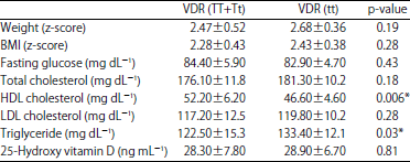
Demographic and anthropometric data of studied children are demonstrated in Table 1. Obese children had significant elevation of total cholesterol, LDL cholesterol, triglycerides and low HDL cholesterol (p<0.001). Also, obese children had significantly low vitamin D than control group (p = 0.002) as present in Table 2. Regarding genotype distributions, no significant difference between cases and controls was observed in genotype and allele frequencies of VDR-FokI (p = 0.63 and 0.74) and VDR-ApaI (p = 0.82 and 0.64). For the TaqI polymorphism, there were significant differences in genotype frequencies of VDR-TaqI between obesity and control groups (p = 0.039) (OR for ‘tt’ allele = 3.47, 95% CI: 1.1-10.7). The ‘t’ allele distribution in the obesity group was significantly higher than control group (p = 0.003) and the OR for ‘t’ was 2.33 (95% CI: 1.34-4.1) as shown in Table 3. Additionally, in obesity-only children carriers for the common allele for VDR were compared their frequency against the rare allele for all parameters of obesity. Homozygotes of the VDR-TaqI ‘t’ variant were associated with increased triglyceride levels (p = 0.03) and HDL levels (p = 0.006) compared with heterozygotes and homozygotes of the ‘T’ allele in obesity subjects (Table 4).
DISCUSSION
Despite growing recognition of the problem, the obesity epidemic continues and obesity rates are increasing around the world24.
| Table 1: | Demographic and anthropometric parameter of the studied groups |
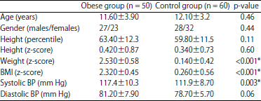 | |
| *Significant | |
| Table 2: | Fasting blood glucose, lipid profile and vitamin D levels of studied groups |
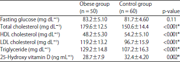 | |
| *Significant | |
| Table 3: | Distribution of vitamin D gene polymorphism in studied groups |
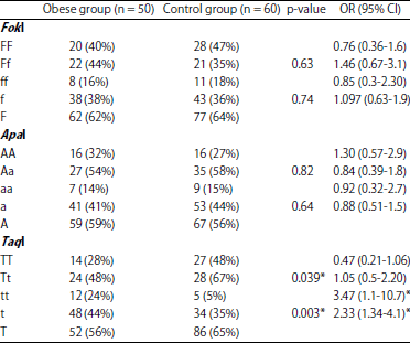 | |
| *Significant | |
| Table 4: | Subject’s anthropometric and laboratory profile according to TaqI gene among obese children |
 | |
| *Significant | |
Therefore, it is widely hoped that the identification of the genetic factors underlying the heritable risk of obesity will contribute to our basic knowledge of the biology of energy balance and might even highlight molecules and pathways that can be targeted for human intervention16.
The VDR genes have been suggested to be potential key players in the pathogenetic mechanism of obesity25. Different ethnic gene and allele variations are in different frequencies, studies have revealed that VDR polymorphisms across ethnics were correlated with different incidences of many diseases. Thus, it was necessary to investigate the possible association between known polymorphisms in VDR genes and the development of obesity in Egyptian children.
Obesity is usually correlated with the higher prevalence of hypovitaminosis or the lower circulating 25(OH) D level in both pediatric as well as adult populations26. Association between hypovitaminosis D and obesity has been also confirmed in the present study. Similarly, a recent Egyptian study reported that the prevalence of 25(OH) D deficiency was higher in the obese group (29.9 ng mL–1) than in the control group (39.7 ng mL–1) with significant difference27. The inverse relationship between obesity and serum 25(OH) D concentrations may have several explanations, including deposition of vitamin D in body fat compartments, reduced release of vitamin D into systemic circulation and low exposure to sun light28. Until now, data regarding the role of vitamin D in obesity are inconclusive29.
The present study found a strong association between VDR-TaqI ‘t’ allele and obesity. This VDR polymorphism on itself or acting as an association signal for a nearby mutation due to linkage disequilibrium, as it happens to be the case for a microsatellite repeat marker in VDR’s 3 UTR30 could affect VDR’s mRNA stability leading to an alteration in its protein expression levels16. Interestingly, when human VDR was over-expressed in the adipocytes of transgenic mice, this resulted ina significant decrease in energy expenditure and induction of obesity30.
In children, there are few studies which investigated the role of VDR polymorphism relation with obesity. Ferrarezi et al.31 reported association between VDR-BsmI polymorphism and height in a cohort of obese children and adolescents; however, there was no significant association between VDR-TaqI and VDR-ApaI.
In a recent study from Kingdom of Saudi Arabia; their data indicated that polymorphisms affecting the vitamin D/VDR axis play a role in obesity that is associated with an ongoing degree of inflammation, possibly resulting from alterations of gut permeability and microbial translocation. The associations with obesity were positive for all studied polymorphisms including BsmI, ApaI and TaqI18.
In contrast, a Polish study reported that there was no statistically significant differences were noted for weight, height and BMI depending on four VDR genotypes (BsmI, FokI, ApaI and TaqI) at all three analyzed loci32.
The influence of VDR polymorphisms on somatic development is one of many genetic factors determining growth and development as well as the involution processes. Moreover, it depends on many environmental factors (geographical, social, economic and cultural). Therefore, the results obtained in various populations are different.
Several studies confirmed the same association in adult’s studies. The association between vitamin D receptor gene polymorphism (TaqI) and obesity was confirmed recently in Chinese population17. These results consistent with other studies in which the TaqI polymorphism presented a significantly higher weight and BMI33,34.
Moreover, homozygotes of the VDR-TaqI ‘t’ allele were associated with increased triglyceride levels and decreased HDL levels, something that suggests an exacerbation of lipid accumulation and triggering of atherosclerosis. Similar results are found also in a Greek study16.
CONCLUSION AND LIMITATION
The present study suggests that TaqI of VDR polymorphisms is associated with obesity in a sample of Egyptian children. The genotype ‘tt’ and allele ‘t’ of TaqI may be potential predictors related to obesity. These results could help the definition of VDR fingerprints that predict an increased risk of developing obesity and might contribute to the identification of novel therapeutic strategies for this metabolic condition.
Despite the significance of our findings, these results should be taken cautiously as our study had some limitations, being mainly the relatively small sample size of obesity as the case-control design that may have allowed some elusive misclassification of controls. Further replication of our findings in larger independent cohorts could overcome these limitations and provide sufficient power to test for gene-gene and gene-diet interactions in order to disentangle the molecular basis of obesity.
REFERENCES
- Ng, M., T. Fleming, M. Robinson, B. Thomson and N. Graetz et al., 2014. Global, regional and national prevalence of overweight and obesity in children and adults during 1980-2013: A systematic analysis for the Global burden of disease study 2013. Lancet, 384: 766-781.
CrossRefDirect Link - Kelishadi, R, S.D. de Ferranti, R. Majdzadeh, J.A. O'Dea, A.K. Gupta and K. Adeli, 2013. Childhood obesity: Today and tomorrow's health challenge. J. Obesity.
CrossRefDirect Link - Dubois, L., K.O. Kyvik, M. Girard, F. Tatone-Tokuda and D. Perusse et al., 2012. Genetic and environmental contributions to weight, height and BMI from birth to 19 years of age: An international study of over 12,000 twin pairs. PLoS ONE, Vol. 7.
CrossRefDirect Link - Lee, Y.S., 2013. Genetics of nonsyndromic obesity. Curr. Opin. Pediatr., 25: 666-673.
CrossRefDirect Link - Rosen, C.J., J.S. Adams, D.D. Bikle, D.M. Black and M.B. Demay et al., 2012. The nonskeletal effects of vitamin D: An Endocrine society scientific statement. Endocr. Rev., 33: 456-492.
CrossRefDirect Link - Hossein-Nezhad, A. and M.F. Holick, 2013. Vitamin D for health: A global perspective. Mayo Clinic Proc., 88: 720-755.
CrossRefDirect Link - Bouillon, R., G. Carmeliet, L. Lieben, M. Watanabe and A. Perino et al., 2014. Vitamin D and energy homeostasis-of mice and men. Nat. Rev. Endocrinol., 10: 79-87.
CrossRefDirect Link - Awad, A.B., L. Alappat and M. Valerio, 2012. Vitamin D and metabolic syndrome risk factors: Evidence and mechanisms. Crit. Rev. Food Sci. Nutr., 52: 103-112.
CrossRefDirect Link - Taymans, S.E., S. Pack, E. Pak, Z. Orban, J. Barsony, Z. Zhuang and C.A. Stratakis, 1999. The human Vitamin D Receptor gene (VDR) is localized to region 12cen-q12 by fluorescent in situ hybridization and radiation hybrid mapping: Genetic and physical VDR map. J. Bone Mineral Res., 14: 1163-1166.
CrossRefDirect Link - Grundberg, E., H. Brandstrom, E.L. Ribom, O. Ljunggren, H. Mallmin and A. Kindmark, 2004. Genetic variation in the human vitamin D receptor is associated with muscle strength, fat mass and body weight in Swedish women. Eur. J. Endocrinol., 150: 323-328.
CrossRefDirect Link - Jain, R., P.R. von Hurst, W. Stonehouse, D.R. Love, C.M. Higgins and J. Coad, 2012. Association of vitamin D receptor gene polymorphisms with insulin resistance and response to vitamin D. Metab. Clin. Exp., 61: 293-301.
CrossRefDirect Link - McDermott, M.F., A. Ramachandran, B.W. Ogunkolade, E. Aganna and D. Curtis et al., 1997. Allelic variation in the vitamin D receptor influences susceptibility to IDDM in Indian Asians. Diabetologia, 40: 971-975.
CrossRefDirect Link - Oh, J.Y. and E. Barrett-Connor, 2002. Association between vitamin D receptor polymorphism and type 2 diabetes or metabolic syndrome in community-dwelling older adults: The Rancho Bernardo study. Metabolism, 51: 356-359.
CrossRefDirect Link - Wong, K.E., J. Kong, W. Zhang, F.L. Szeto and H. Ye et al., 2011. Targeted expression of human vitamin D receptor in adipocytes decreases energy expenditure and induces obesity in mice. J. Biol. Chem., 286: 33804-33810.
CrossRefDirect Link - Vasilopoulos, Y., T. Sarafidou, K. Kotsa, M. Papadimitriou and Y. Goutzelas et al., 2013. VDR TaqI is associated with obesity in the Greek population. Gene, 512: 237-239.
CrossRefDirect Link - Fan, H.R., L.Q. Lin, H. Ma, Y. Li and C.H. Sun, 2015. Association between vitamin D receptor gene polymorphism (TaqI) and obesity in Chinese population. J. Genet., 94: 473-478.
CrossRefDirect Link - Al-Daghri, N.M., F.R. Guerini, O.S. Al-Attas, M.S. Alokail and K.M. Alkharfy et al., 2014. Vitamin D receptor gene polymorphisms are associated with obesity and inflammosome activity. PLoS ONE, Vol. 9.
CrossRefDirect Link - Ezzat, A.M., M.F. EL Gendy, D.R. Soliman and H.S.A.A. Abou Ghazy, 2011. Body mass index as an assessment tool for overweight and obesity in school children in El-Qalubia governorate. J. Am. Sci., 7: 240-250.
Direct Link - Kuczmarski, R.J., C.L. Ogden, S.S. Guo, L.M. Grummer-Strawn and K.M. Flegal et al., 2002. 2002 CDC growth charts for the United States: Methods and development. Vital Health Stat., 11: 1-190.
PubMedDirect Link - CDC., 2011. CDC grand rounds: Childhood obesity in the United States. Morbidity Mortality Weekly Rep., 60: 42-46.
PubMedDirect Link - Miller, S.A., D.D. Dykes and H.F. Polesky, 1988. A simple salting out procedure for extracting DNA from human nucleated cells. Nucleic Acids Res., 16: 1215-1215.
PubMedDirect Link - Mitchell, N., V. Catenacci, H.R. Wyatt and J.O. Hill, 2011. Obesity: Overview of an epidemic. Psychiatr. Clin. North Am., 34: 717-732.
CrossRefDirect Link - Perusse, L. and C. Bouchard, 2000. Gene-diet interactions in obesity. Am. J. Clin. Nutr., 72: 1285s-1290s.
Direct Link - Lagunova, Z., A.C. Porojnicu, R. Vieth, F.A. Lindberg, S. Hexeberg and J. Moan, 2011. Serum 25-hydroxyvitamin D is a predictor of serum 1,25-dihydroxyvitamin D in overweight and obese patients. J. Nutr., 141: 112-117.
CrossRefPubMedDirect Link - Hassan, N.E., S.A. El-Masry, R.A. El Banna, M.M. Abu Shady and M. Al-Tohamy et al., 2014. 25-hydroxy vitamin D, adiponectin levels and cardiometabolic risk factors in a sample of obese children. Macedonian J. Med. Sci., 7: 562-566.
CrossRefDirect Link - Wortsman, J., L.Y. Matsuoka, T.C. Chen, Z. Lu and M.F. Holick, 2000. Decreased bioavailability of vitamin D in obesity. Am. J. Clin. Nutr., 72: 690-693.
Direct Link - Ross, A.C., J.E. Manson, S.A. Abrams, J.F. Aloia and P.M. Brannon et al., 2011. The 2011 report on dietary reference intakes for calcium and vitamin D from the Institute of Medicine: What clinicians need to know. J. Clin. Endocrinol. Metab., 96: 53-58.
CrossRefDirect Link - Ingles, S.A., R.W. Haile, B.E. Henderson, L.N. Kolonel and G. Nakaichi et al., 1997. Strength of linkage disequilibrium between two vitamin D receptor markers in five ethnic groups: Implications for association studies. Cancer Epidemiol. Biomarkers Prev., 6: 93-98.
Direct Link - Ferrarezi, D.A.F., N. Bellili-Munoz, C. Nicolau, N. Cheurfa and I.C. Guazzelli et al., 2012. Allelic variations in the vitamin D receptor gene, insulin secretion and parents' heights are independently associated with height in obese children and adolescents. Metabolism, 61: 1413-1421.
CrossRefDirect Link - Jakubowska-Pietkiewicz, E., I. Klich, W. Fendler, W. Mlynarski and D. Chlebna-Sokol, 2013. Effect of Vitamin D Receptor gene (VDR) polymorphism on body height in children-own experience. Postepy Higieny Medycyny Doswiadczalnej, 67: 873-878.
PubMedDirect Link - Binh, T.Q., Y. Nakahori, V.T.T. Hien, N.C. Khan, N.T. Lam, L.B. Mai and S. Yamamoto, 2011. Correlations between genetic variance and adiposity measures and gene × gene interactions for obesity in postmenopausal Vietnamese women. J. Genet., 90: 1-9.
CrossRefDirect Link - Ye, W.Z., A.F. Reis, D. Dubois-Laforgue, C. Bellanne-Chantelot, J. Timsit and G. Velho, 2001. Vitamin D receptor gene polymorphisms are associated with obesity in type 2 diabetic subjects with early age of onset. Eur. J. Endocrinol., 145: 181-186.
CrossRefDirect Link