
Arash Omidi
Department of Animal Sciences, Faculty of Agriculture, University of Birjand, P.O. Box 91775-331, Birjand, Iran
Asian Journal of Animal and Veterinary Advances
Year: 2008 | Volume: 3 | Issue: 5 | Page No.: 381-385


ABSTRACT
The aim of this study was to introduce abscess on left thoracic wall of cattle as less common complication of traumatic reticulitis and finding the best way for diagnosing them. During two years, among 75 cases confirmed suffering from traumatic reticulitis, five cows with thoracic abscess were examined clinically, radiographically and ultrasonographically. Clinical signs observed included anorexia, pyrexia, abdominal pain and weight loss. All of the cases had a big abscess in the back portion of left humerus on the thoracic wall. In every case, the diagnosis was confirmed by the centesis and aspiration of the abscess. In all cows, radiographic findings revealed a metal foreign body in the reticulum penetrating it. Ultrasonography revealed a large reticular abscess with a well-developed capsule that appeared as echogenic deposits that were sometimes accompanied by hypoechogenic fluid. The abscess was elongated towards the left thoracic wall and appeared in the posterior portion of the left humerus. The abscess was incised and drained from body surface and the reticulum during a rumenotomy. After the abscess drainage and treatment with antibiotics, all cows recovered and became healthy. It is concluded that foreign bodies should be considered in the differential diagnosis of localised swelling on the thoracic wall and the best way for diagnosing them is a combination of clinical findings, laboratory testing, ultrasonography and radiography.
PDF Abstract XML References Citation
How to cite this article
Arash Omidi, 2008. Less Common Complication of Traumatic Reticulitis in Cattle: Abscess on Left Thoracic Wall. Asian Journal of Animal and Veterinary Advances, 3: 381-385.
DOI: 10.3923/ajava.2008.381.385
URL: https://scialert.net/abstract/?doi=ajava.2008.381.385
DOI: 10.3923/ajava.2008.381.385
URL: https://scialert.net/abstract/?doi=ajava.2008.381.385
INTRODUCTION
Traumatic reticuloperitonitis, or TRP, is a relatively common disease in adult cattle caused by the ingestion and migration of a foreign body in the reticulum. Cattle are more likely to ingest foreign bodies than small ruminants since they do not use their lips for prehension and are more likely to eat a chopped feed. The typical foreign body is a metallic object, such as a piece of wire or a nail (Rebhun, 1995). Due to the perforation of the reticulum by sharp foreign bodies that have been ingested, TRP has been a concern of veterinarians for many years. The prevalence of traumatic reticulitis in adult dairy cattle has been attributed to management practices and the lack of discriminatory dietary habits of cattle (Roth and King, 1991). The classic signs associated with TRP are consistent with an acute, localized peritonitis and include anorexia, fever, tachypnea and an arched stance with abducted elbows (indicating cranial abdominal pain). If the foreign body has penetrated the diaphragm and pericardium, affected cattle also can have muffled heart sounds, jugular pulses and brisket edema secondary to congestive heart failure caused by pericarditis. However, not all cattle develop acute peritonitis; a significant population of affected cattle develops chronic or subclinical TRP that is not as easily diagnosed as acute TRP (Hawkins, 2002). Although it has been suggested that traumatic reticulitis should be considered in any bovine animal suffering from indigestion, the variability of the clinical signs has also been stressed (Roth and King, 1991). Reticular abscesses are
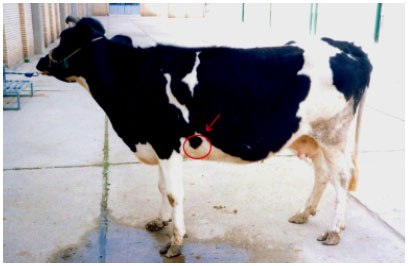 | |
| Fig. 1: | Abscess position (arrow) |
a common complication of TRP. In addition, if the foreign body migrates through the diaphragm and into the pericardium, it can cause septic pericarditis and result in a subsequent congestive heart failure. Some wires have an unusually pattern of movement in some cases and make less common complications include reticular fistulation, vagal indigestion and diaphragmatic hernia (Rebhun, 1995; Ibrahim and Gohar, 1988). There are few references to the cliical diagnosis and management of thoracic abscesses in cattle (Braun et al., 2007, 1998, 1993a). The purpose of this study was to introduce unusual cases of traumatic reticulitis which appears as abscess on the left thoracic walls of cows (Fig. 1) and finding the best practical application in routine diagnostic tests.
MATERIALS AND METHODS
Over a 2 year period, abscess on thoracic wall was diagnosed in 5 cattle among 75 cases confirmed suffering from traumatic reticulitis in veterinary hospital of Shahid Chamran University (2004-2006), five Holstein-Frisian dairy cattle have had an abscess on the back portion of the left humerus which were examined clinically, haematologically, radiographically and ultrasonographically. All of the five cattle were female; the age of them ranged from 4 to 7 years with an average age of 4.8 years ± 0.83 SD.
In every case, the diagnosis was confirmed by the centesis and aspiration of the abscess, which yielded purulent material. The areas over the reticulum and the left and right sides of the thorax up to the level of the elbow joints were clipped. The remaining hair was removed with depilatory cream, transmission gel was applied and the cows were examined ultrasonographically with a 3.5 MHZ linear array transducer with a radius of 40 mm and then, the morphological changes in the region of the abscess were examined in the cows. A radiograph of the reticulum was also taken (Misk and Semieka, 2001). Blood samples were collected from each animal for hematological examination. All owners having animals with abscesses accepted treatment for their animals. The findings of the clinical and radiographic diagnosis were compared with those of ultrasonography.
RESULTS AND DISCUSSION
All cattle all had clinical signs including chronic indigestion, pyrexia (mild to moderate), reduced ruminal motility (stasis until 2 contractions every 5 min) and weight loss. All of these five cattle have had a big abscess in the back portion of the left humerus on the thoracic wall (Fig. 2). The diameters
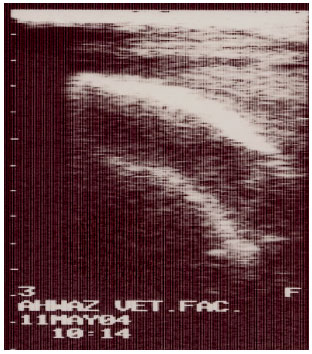 | |
| Fig. 2: | Ultrasonography revealed a large reticular abscess in the posterior part of the left humerus of cows, the Hyperechogenic line is related to the reticular foreign body |
of the abscesses were 7-10 cm in 2 animals and 15-21 cm in the other three. Various pain tests, such as the back grip test, pole test and pain percussion on the xiphoid region, were positive in all the cows. A hematological examination revealed anemia, increased concentrations of plasma protein and fibrinogen in all of the cases. In two cases, a significant increase in leukocyte count was noticed. A radiographically identified penetrating reticular foreign body is a near-certain cause of traumatic reticulitis, parareticular abscessation, or peritonitis (Dor et al., 2007). An extrareticular wire or nail is the most likely cause of reticulitis or peritonitis in an animal with compatible clinical signs. An immobile reticular foreign body may be trapped in the reticular mucosa, penetrating a mucosal fold (but not the reticular wall), or piercing the wall of the reticulum (Farrow, 1999). In all cows, radiographic findings revealed a metal foreign body in the reticulum penetrating it. Radiography was best suited for visualization of metallic foreign bodies in and outside the reticulum and the position of the foreign body was not a reliable indicator of the condition. Radiography is the best method for visualizing them and for obtaining accurate information about their position and nature (Braun et al., 1993a). All of these foreign bodies revealed a large reticular abscess with a well-developed capsule appeared as echogenic deposits that sometimes accompanied by hypoechogenic fluid. Abscesses had an echogenic capsule with a hypoechogenic center which was same as findings of other researchers (Braun et al., 1993b). Ultrasonographic examination revealed the presence of fibrinous changes or abscesses that could not be visualized by radiography. The major advantage of ultrasonography overcomes the problem of locating the lesion but also its size and extent. In contrast, ultrasonography failed to identify any metallic objects including magnets. Indeed, ultrasound was a useful diagnostic tool to help retrieve a fluid sample for culture and sensitivity, monitor the progression of the lesion and to guide surgical debridement (Mohamed and Oikawa, 2007; Braun et al., 1998). The abscess was elongated towards the left thoracic wall and appeared in the posterior portion of the left humerus. In one case, the tip of wire was seen on the abscess surface. In other cases, the skin was intact. In all the animals, after sterilization by a standard surgical disinfection technique, the abscess was incised and drained from the body surface and the reticulum during the left flank exploratory laparotomy (McGavin and Morril, 1976). After the abscess drainage and a medical treatment that involves the administration of antibacterial (Penicillin) and supportive therapy (calcium borogluconate was administered for correcting ruminal stasis), all cows recovered their health (Radostits et al., 2007). Because of the non-specific clinical signs, the possible diagnosis of abdominal and thoracic abscesses is frequently not taken into consideration. Hence, thoracic abscesses may be more common than is currently appreciated as little accurate information on the prevalence or incidence is available (Mohamed and Oikawa, 2007). Ultrasonography alone cannot ensure an accurate diagnosis and a differential diagnosis must include neoplasia, cysts (Mohamed and Oikawa, 2007) or granulomatous lesions (Yeruham et al., 2004). Radiography plays an important role in the diagnosis of many thoracic disorders (Misk and Semieka, 2001).
The results of the present investigation illustrate the effectiveness of the clinical findings, laboratory testing, ultrasonography and radiography combination. As a conclusion combining these various techniques with each other have a large practical application in the diagnosis and treatment of the traumatic reticulitis complications. Foreign bodies should be considered in the differential diagnosis of localised swelling on the thoracic wall (Dor et al., 2007; Mohamed and Oikawa, 2007; Ihsan et al., 2003; Braun et al., 1999).
ACKNOWLEDGMENTS
This research was carried out by financial support of the University of Birjand. I would like to express my special thanks to Dr. A.R. Ghadiri and Dr. F.S. Afshar (School of Veterinary Medicine, Shaid Chamran University of Ahwaz) for ultrasonography, radiography and surgery of the cases and to Dr. Wasim Tawbi for his constructive suggestions to write up the manuscript.
REFERENCES
- Braun, U., M. Fluckiger and F. Nageli, 1993. Radiography as an aid in the diagnosis of traumatic reticuloperitonitis in cattle. Vet. Rec., 132: 103-109.
PubMedDirect Link - Braun, U., M. Gotez and O. Marmier, 1993. Ultrasonographic findings in cows with traumatic reticuloperitonitis. Vet. Rec., 133: 416-422.
PubMedDirect Link - Braun, U., U. Iselin, C. Lischer and E. Fluri, 1998. Ultrasonographic findings in five cows before and after treatment of reticular abscesses. Vet. Rec., 142: 184-189.
CrossRefPubMedDirect Link - Braun, U., C. Lischer, U. Koller, R. Mller and U. Geissbhler, 1999. Imaging findings in a cow with a retropharyngeally displaced magnet. Vet. Radiol. Ultrasound, 40: 162-163.
CrossRefDirect Link - Braun, U., B. Lejeune, G. Schweizer, M. Puorger and F. Ehrensperger, 2007. Clinical findings in 28 cattle with traumatic pericarditis. Vet. Rec., 161: 558-563.
Direct Link - Dor, E., G. Fecteau, P. Hlie and D. Francoz, 2007. Liver abscesses in Holstein dairy cattle: 18 cases (1992-2003). J. Vet. Intern. Med., 21: 853-856.
Direct Link - Farrow, C., 1999. Reticular foreign bodies. Causative or coincidence? Vet. Clin. North Am. Food Anim. Pract., 15: 397-408.
PubMedDirect Link - Ibrahim, I. and H. Gohar, 1988. Unusual pathway of a penetrating reticular foreign body (wire) in a buffalo (clinical report). Egypt. J. Vet. Sci., 1: 85-87.
Direct Link - Ihsan, K., I. Alptekin, N. Atasoy, A. Cinar, N. Donmez and E. Ceylan, 2003. Pseudopericarditis in a cow caused by theileriosis - A case report. Vet. Arhive., 73: 111-117.
Direct Link - McGavin, M.D. and J.C. Morril, 1976. Dissection technique for examination of the bovine ruminoreticulum. J. Anim. Sci., 42: 535-538.
Direct Link - Misk, N.A. and M.A. Semieka, 2001. The radiographic appearance of reticular diaphragmatic herniation and traumatic pericarditis in buffaloes and cattle. Vet. Radiol. Ultrasoun., 42: 426-430.
Direct Link - Mohamed, T. and S. Oikawa, 2007. Ultrasonographic characteristics of abdominal and thoracic abscesses in cattle and buffaloes. J. Vet. Med. Ser. A, 54: 512-517.
CrossRefPubMedDirect Link - Roth, l. and J.M. King, 1991. Traumatic reticulitis in cattle: A review of 60 fatal cases. J. Vet. Diag. Invest., 3: 52-54.
Direct Link - Yeruham, I., S. Friedman, S. Perl, D. Elad, Y. Berkovich and Y. Kalgard, 2004. A herd level analysis of a Corynebacterium pseudotuberculosis outbreak in a dairy cattle herd. Vet. Dermatol., 15: 315-320.
Direct Link