
R.M. Dharmadasa
Herbal Technology Section, Industrial Technology Institute, 363, Bauddhaloka Mawatha, Colombo 7, Sri Lanka
G.A.S. Premakumara
Herbal Technology Section, Industrial Technology Institute, 363, Bauddhaloka Mawatha, Colombo 7, Sri Lanka
P.L. Hettiarachchi
Department of Botany, University of Sri Jayewardenepura, Gangodawila, Nugegoda, Sri Lanka
W.D. Ratnasooriya
Department of Zoology, University of Colombo, Sri Lanka
Research Journal of Medicinal Plants
Year: 2012 | Volume: 6 | Issue: 3 | Page No.: 267-273


ABSTRACT
Munronia pinnata (Wall.) Theob. (Meliaceae) is therapeutically important medicinal plant used for the control of malaria in traditional medicine in Sri Lanka. This study described the cytotoxic potential of five morphotypes and antimalarial activity of the highest cytotoxic morphotype of M. pinnata using brine shrimp toxicity assay and Plasmodium yoelii murin model, respectively. Four concentrations of (0.1, 1, 5 and 10 mg mL-1) aqueous extracts of stem, roots and leaves of five morphotypes of M. pinnata were screened for cytotoxcity using brine shrimp assay. Either, three doses (3200, 1600, 800 mg kg-1) of the whole plant aqueous extract of the highest cytotoxic morphotype of M. pinnata (WPAE) or vehicle or quinine were orally administered for different groups of mice (n = 6) inoculated with P. yoelii murine model. Antimalarial activity was examined in terms of schizonticidal activity and chemo suppression. Overt signs of toxicity, body weight, rectal temperature, stress and aversive behavior were observed with long term oral administration of high dose of WPAE (n = 10). All parts of five morphotypes of M. pinnata showed potent cytotoxic activity. The order of potency was roots>stems>leaves. The highest cytotoxicity exhibited by morphotype collected from Nilgala forest reserve (MGNG3). The high dose of WPAE of MGNG3 has potent oral antimalarial activity. No signs of overt toxicity, signs of stress and aversive behaviour observed during long term toxicological studies. This study showed, for the first time, that all morphotypes of M. pinnata has marked cytotoxcity and morphotype MGNG3 has potent antimalarial activity as claimed by Sri Lankan traditional practitioners.
PDF Abstract XML References Citation
Received: October 29, 2011;
Accepted: November 17, 2011;
Published: January 10, 2012
How to cite this article
R.M. Dharmadasa, G.A.S. Premakumara, P.L. Hettiarachchi and W.D. Ratnasooriya, 2012. Cytotoxcity and in vivo Antimalarial Activity of Aqueous Whole Plant Extract of Munronia pinnata (Wall.) Theob. (Meliaceae) in Mice. Research Journal of Medicinal Plants, 6: 267-273.
URL: https://scialert.net/abstract/?doi=rjmp.2012.267.273
URL: https://scialert.net/abstract/?doi=rjmp.2012.267.273
INTRODUCTION
Malaria is one of the most dangerous parasitic diseases and major global health problem affecting over one hundred countries particularly in the last two decades mainly in African and Asian countries (Guerin et al., 2002). Vivas et al. (2005) pointed out that over 300-500 million new clinical cases of malaria are reported and kill one to two million children in each year. The increased incidence of malaria in recent years has been attributed to the development of resistance of the malarial protozoa to existing antimalarial drugs like chloroquine (Peters, 1998; Kaur et al., 2009). At present several antimalarial drugs such as artesunate, artemether, quinine, artimisine and chloroquinine are used to treat malaria. However, rapid development of resistance by malarial protozoa to the existing drugs, created an urgent necessity for new antimalarial drugs (Zhang et al., 2004).
Historically malaria and many other dangerous diseases have been controlled by using natural origins such as plants, bacteria, fungi and marine sources (Phillipson and Wright, 1991). Moreover, approximately 80% of the world’s population still depends on traditional medicine as a source of treatment for their primary health care needs (Phillipson and Wright, 1991; WHO, 2002). Further, 78% of the new drugs approved by the FDA between 1983 and 1994 were derived from natural products or semi-synthetically obtained from natural biota (Cragg et al., 1997). Meanwhile, some of the prominent effective antimalarial drugs such as quinine and artemisinin have been obtained from plants (Basco et al., 1994). Further, importance of discovery of newer, effective, safe drugs from medicinal plants used in traditional systems of medicine have been emphasized by many workers (Sarwar et al., 2011; Gansane et al., 2010). Therefore, plants with traditional claims play an important role in discovery of newer effective, safe, alternative antimalarial drugs.
M. pinnata (Wall) Theob. (Meliaceae) is a therapeutically important plant species which is widely used in traditional and folklore medicine and also by Sri Lankan aborigine populations for antimalarial treatments (Jayaweera, 1982). It has 5 morphotypes in different localities in the country: having 3,5,7,9 and 11 leaflet bearing compound leaves of which three leaflets type is widely used therapeutically. In Sri Lankan traditional and folklore medicine the entire plant is used in more than 32 recipes for the treatment of malaria, recurrent fever, dysentery and purification of blood (Annonymous, 1979; Jayaweera, 1982). Recent scientific investigations conducted by MacKinnon et al. (1997), Roy and Saraf (2006) revealed that plants of family Meliaceae contain range of biologically active compounds which are responsible for an array of biological activities such as insecticidal, insect antifeedant and insect growth regulation as well as a number of other pharmacological activities on humans including antibacterial, antifungal, antimalarial, anticancer and antiviral. However, scientific investigation/s on cytotoxcity of M. pinnata is yet to be reported. Further, traditional and folklore claims of antimalarial activity of M. pinnata is not scientifically tested and validated. The aim of this study was to examine the cytotoxic and antimalarial activity of M. pinnata using brine shrimp toxicity assay and P. yoelii murin model, respectively.
MATERIALS AND METHODS
Collection of plant material: This study was conducted from June 2009 to August 2010. M. pinnata plants were collected from different agro-climatic regions of Sri Lanka and identified by comparing with authentic herbarium specimens deposited at Royal Botanical Garden, Peradeniya, Sri Lanka. Morphotypes codes with four letters and a number were assigned to each morphotype by indicating collected district, name of the exact location of collection and the number of leaflets in each morphotype: For APRG5 (Anuradhapura district, Ritigala area bearing five leaflets), BDHM3 (Badulla district, Haldummulla area bearing three leaflets), GPWP3 (Gampaha district, Warakapola area bearing three leaflets), MGMD3 (Moneragala district, Madulla area with three leaflets) and MGNG3 (Moneragala district, Nilgala area with three leaflets). Herbarium specimens were prepared and deposited at the institutional herbarium (HTS-1-HTS-5).
Preparation of crude extracts: The plants were separated into roots, stems and leaves and dried in shade (30±2°C) for three days. Dried parts were powdered using an electrical grinder (Janke and Kundel, Staufen, Germany) Sixty gram of powdered material of each part was extracted into 1920 mL of water under constant heat for 4 h until the volume of the solution was reduced to 1/8 (240 mL) of the initial volume as described in traditional medicine in Sri Lanka. Extracts were filtered through Whatman No.1 filter paper and filtrates were separately freeze-dried (yield 10 % w/w) and extracts were made into 0.1, 1, 5 and 10 mg mL-1 concentrations were made using artificial saline water (35 ppt). The same procedure was used to prepare whole plant aqueous extracts (WPAE) of morphotype (MGNG3) of M. pinnata having the highest brine shrimp toxicity. Freeze dried product of WPAE was dissolved in water in order to prepare the extracts for antimalarial assay.
Brine shrimp toxicity assay: Brine shrimp toxicity assay was performed as described by Michael et al. (1956) with slight modifications. Artemia salina eggs purchased from a local aquarium were incubated in 100 mL of artificial sea water (pH 7-8 and salinity 35 ppt) under artificial light at 28°C. After incubation for 24 h, the nauplii were collected with a Pasteur pipette and kept for an additional 24 h under the same conditions to reach the meta nauplii stage. After 48 h of hatching, the shrimp larvae were transferred to 18 well plates containing 1 mL of aerated artificial sea water. The extracts of 4 different concentrations (0.1, 1, 5 and 10 mg mL-1) were added to different wells and shaken lightly to ensure a homogeneous test solution and left at room temperature (28°C). Artificial seawater was used as the control. The number of dead larvae in each well after 24 h period was determined with the aid of a light microscope (Olympus, CX31 and Japan) at 40x and was used as a measure of toxicity of the extracts.
In vivo antimalarial activity
Experimental animals: Healthy, adult male C57BL/6JNcl mice weighing 30-41 g (9 week old), purchased from Medical Research Institute, Colombo, Sri Lanka were used in this experiment. All animals were reared in plastic cages in the animal house, Department of Zoology, University of Colombo, under standard conditions [Temperature 28±2°C; photoperiod, approximately 12 h natural light per day, Relative Humidity 50-55%). The animals were fed with pelleted food (Master Feed Ltd. Colombo, Sri Lanka) and clear drinking water ad libitum. Except at the time of experimental procedure the animals were carefully handled only during cage cleaning. The study was conducted in accordance with the internationally accepted guidelines for laboratory animal use and care and guidelines and rules of the Faculty of Science, University of Colombo Sri Lanka. All mice were acclimatized for 7 days before incorporating into the experiment.
Parasite isolates: Chloroquine sensitive, 17XL (lethal strain) Plasmodium yoelii was used to assess the in vivo antimalarial activity of WPAE. The parasite strain was maintained by serial passage of blood from mouse to mouse.
Parasitic inoculation: The inoculum consisted of 107 P. yoelii parasitized red blood cells (RBCs)/mL of blood. This was prepared by determining both the percentage parasitaemia and the erythrocyte counts of the donor mouse and diluting the blood with isotonic saline.
Evaluation of blood schizonticidal activity on an early infection: The 4-day suppressive assay was conducted to evaluate the blood schizonticidal activity of the WPAE. Mice were intraperitonially inoculated with 0.2 mL of saline, infected with P. yoelii (Containing 107RBC) on day zero. The inoculated mice were then randomly put into cages (N = 6/cage). Rectal temperatures and body weight of each animal were recorded before the drug administration. Three doses of the WPAE (3200 (T1), 1600 (T2), 800 mg kg-1 (T3), 10 mg kg-1 quinine (State Pharmaceutical Corporation, Colombo, Sri Lanka) as the positive control (PC) and distilled water as the negative control (C) were orally administrated to each group (N = 6/cage) for 4 days. From day 3, blood was obtained from the tail of each mouse under aseptic conditions and blood smears were made on clean glass slides. The number of parasitized RBC was determined by counting at least 3000 erythrocytes in random fields under oil immersion. The average percentage of chemosuppresion was calculated as:
Mice used for the 4-day suppressive assay were further observed daily for eight consecutive days and the mean survival time (days), body weight (g) and rectal temperature (°C) for each mouse were recorded.
Toxicological studies: The study was carried out to determine overt signs of toxicity of WPAE in mice. Twenty mice were assigned into two groups (N = 10/group). Body weight and rectal temperature of each animal were recorded. One group was administered with 3200 mg kg-1 (500 μL of extract) dose of WPAE and the other group received same volume of distilled water (control). The general behaviour of mice was observed continuously for one hour after the treatment and then intermittently for 4 h. The mice were further observed up to 90 days for gross changes in behaviour and manifestations of any overt signs of toxicity (increased motor activity, tremors, convulsions, watery diarrhea, salivation and fur yellowing, postural abnormalities and behavioral changes), signs of stress (fur erection, exophthalmia) and aversive behaviour (biting and scratching behavior, licking of tail, paw and penis, intense grooming behavior or vocalization). Body weights were recorded before administration of the extract in each day.
Statistical analysis: Mortality of brine shrimp in different concentrations was counted after 24 h. Each treatment was replicated four times. LC50 values were generated by probit analysis using SPSS version 10 statistical packages and presented as LC50±SE with 95% confidential level. Results of the antimalarial activity were analyzed by General Linear Model (GLM) of ANOVA test and mean comparison followed by Duncan’s Multiple Range Test (DMRT).
RESULTS
Brine shrimp toxicity assay: As shown in Table 1 leaf stem and root extracts of all morphotypes of M. pinnata exhibited cytotoxcity. Moreover, leaf, stem and root extracts of morphotype MGNG3 exhibited the highest cytotoxcity. Order of potency was roots>stem>leaf (LC50 values were 0.02±0.00 mg mL-1 0.18±0.05 mg mL-1, 0.18±0.04 mg mL-1, respectively).
Evaluation of blood schizonticidal activity on an early infection: As shown in Table 2, the high dose (3200 mg kg-1 WPAE (T1) significantly reduced the parasitaemia in the 4-day suppressive assay compared with the control, T2 and T3 at 5% significant level.
| Table 1: | LC50 values for different parts of five morphotypes of Munronia pinnata |
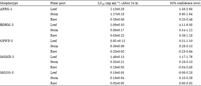 | |
| Results are the mean of 4 replicates. Values are as Mean±SE | |
| Table 2: | In vivo blood schizonticidal activity, chemosuppresion and survival rate in the 4-day suppressive assay |
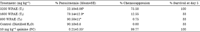 | |
| Values denoted by different letters are significantly different at 5% significant level, WPAEMP = Aqueous Extract of M. pinnata, n = 6 | |
Moreover, the high dose of WPAE produced huge and significant chemosuppresion (75.58%) and 100% survival rate (Table 2). However, the chemosuppression and schizonticidal activity was dose dependent. In contrast, the reference drug, quinine suppressed parasitaemia by almost 100% (Table 2). The survival rates of lower doses and control were poor.
All the mice in T2, T3 and control groups died after six days. Therefore, parasitaemia percentage in T1 and positive control were monitored up to eight days. Average rectal temperatures of mice in control group increased from 36.14±0.17°C to 38.30±0.37°C while it did not show an increase in T1 (36.22±0.17°C-36.87±0.39°C) and positive control (36.12±0.21°C-36.63±0.26°C) groups. However, body weight of mice in control, T2 and T3 group reduced while it was gradually increased in mice in T1 and PC treatments.
Toxicological studies: None of the treated mice died. Further, there were no overt signs of toxicity, stress and aversive behaviours observed in WPAE treated mice as in the control mice just after administration of the extracts and also during whole experimental period.
DISCUSSION
This study examined the cytotoxcity of five morphotypes of M. pinnata and the antimalarial potential of the highest cytotoxic morphotype (MGNG3). Cytotoxic potential was investigated using the brine shrimp toxicity assay which is considered as a simple, low-cost, highly sensitive, efficient rapid, bench top cytotoxcity screening assay which requires only a relatively small amount of sample (2-20 mg). Since it has a good correlation with cytotoxic activity, it has been extensively used as a preliminary tool for screening of cytotoxicity of plant crude extracts and different fractions of crude extracts (Vivas et al., 2005; Kaur et al., 2009; Gilani and Atta-ur-Rahman, 2005; Anwar et al., 2003).
Antimalarial activity was tested using P. yeolii murine model. This is a widely used reliable and sensitive in vivo model to detect the antimalarial activity of potential antimalarial drugs including medicinal plants which are effective against the P. falciparum human parasites (Peters et al., 1975).
Further, the schizonticidal activity and chemosuppresion of WPAE was dose depended. This indicates a genuine therapeutic effect which is possibly receptor mediated. These results directly support the antimalarial potential of M. pinnata claimed by traditional practitioners of Sri Lanka and also show the possibility of developing an active, safe and potent plant based drug for malaria. M. pinnata also exhibited antipyretic properties. Peters (1998), has pointed out the necessity of developing herbal anti-malarial drugs by considering the emergence and spread of drug resistance in the malaria parasite. Further, possible antimalarial activities of Artemisia afra (Mukinda and Syce, 2007), Morinda lucida and Alstonia boonei (Bello et al., 2009; Phellinus linteus (Samchai et al., 2009) have been investigated.
Annonymous (1998) has reported the presence of limonoids and triterpenoids in members of the family Meliaceae which are responsible for an array of biological activities such as antibacterial, antifungal, antimalarial, anticancer, antiviral and a number of other pharmacological activities on human. Presence of triterpens and limonoids in M. henryi, a Munronia species found in China has been reported by Dua et al. (2004) and Qi et al. (2003). Therefore, it is possible that observed antimalarial activity of WPAE mediated via limonoides and terpens either singly or in synergy. Preliminary toxic studies carried out indicated that the extracts appears to be safe and does not induce stress and aversive behavior.
CONCLUSIONS
This study showed for the first time that all morphotypes of M. pinnata has marked cytotoxcity and morphotype MGNG3 has potent antimalarial activity as claimed by Sri Lankan traditional practitioners.
REFERENCES
- Roy, A. and S. Saraf, 2006. Limonoids: Overview of significant bioactive triterpenes distributed in plants kingdom. Biol. Pharmaceut. Bull., 29: 191-201.
Direct Link - Cragg, G.M., D.J. Newman and K.M. Snader, 1997. Natural products in drug discovery and development. J. Nat. Prod., 60: 52-60.
CrossRefPubMedDirect Link - Dua, V.K., V.P. Ojha, R. Roy, B.C. Joshi and N. Velccha et al., 2004. Antimalarial activity of some xanthones isolated from the roots of Andrographis paniculata. J. Ethnopharmacol., 95: 247-251.
Direct Link - Gilani, A.H. and Atta-ur-Rahman, 2005. Trends in ethnopharmacology. J. Ethnopharmacol., 100: 43-49.
CrossRefDirect Link - Kaur, K., M. Jain, T. Kaur and R. Jain, 2009. Antimalarials from nature. Bioorg. Med. Chem., 17: 3229-3256.
CrossRefDirect Link - MacKinnon, S., T. Durst, J.T. Arnason, C. Angerhofer and J. Pezutto et al., 1997. Antimalarial activity of tropical Meliaceae extracts and Gedunin derivatives. J. Nat. Prod., 60: 336-341.
PubMed - Michael, A.S., C.G. Thompson and M. Abramovitz, 1956. Artemia salina as a test organism for bioassay. Science, 123: 464-464.
CrossRefPubMedDirect Link - Mukinda, J.T. and J.A. Syce, 2007. Acute and chronic toxicity of the aqueous extract of Artemisia afra in rodents. J. Ethnopharmacol., 112: 138-144.
CrossRefPubMedDirect Link - Peters, W., 1975. The chemotherapy of rodent malaria, XXII. The value of drug-resistant strains of P. berghei in screening for blood schizontocidal activity. Ann. Trop. Med. Parasitol., 69: 155-171.
PubMedDirect Link - Peters, W., 1998. Drug resistance in malaria parasites of animals and man. Adv. Parasitol., 41: 1-62.
CrossRefPubMedDirect Link - Phillipson, J.D. and C.W. Wright, 1991. Can ethnopharmacology contribute to the development of antimalarial agents? J. Ethnopharmacol., 32: 155-165.
PubMedDirect Link - Qi, S.H., L. Chen, D.G. Wu, Y.B. Ma and X.D. Luo, 2003. Novel tetranortriterpenoid derivatives from Munronia henryi. Tetrahedron, 59: 4193-4199.
CrossRefDirect Link - Vivas, L., A. Easton, H. Kendrick, A. Cameron and J.L. Lavandera et al., 2005. Plasmodium falciparum: Stage specific effects of a selective inhibitor of lactate dehydrogenase. Exp. Parasitol., 111: 105-114.
PubMed - Zhang, H., M. Paguio and P.D. Roepe, 2004. The antimalarial drug resistance protein Plasmodium falciparum chloroquine resistance transporter binds chloroquine. Biochemistry, 43: 8290-8296.
CrossRef - Anwar, S., B. Ahmad, M. Subhan, W. Gul and Nazar-ul-Islam, 2003. Biological and pharmacological properties of Aconitum chasmanthum. J. Biol. Sci., 3: 989-993.
CrossRefDirect Link - Basco, L.K., S. Mitaku, A.L. Skaltsounis, N. Ravelomanantsoa, F. Tillequin, M. Koch and J. Le Bras, 1994. In vitro activities of furoquinoline and acridone alkaloids against Plasmodium falciparum. Antimicrob. Agents Chemother., 38: 1169-1171.
PubMed - Samchai, S., P. Seephonkai, A. Sangdee, A. Puntumchai and U. Klinhom, 2009. Antioxidant, cytotoxic and antimalarial activities from crude extracts of mushroom Phellinus linteus. J. Biol. Sci., 9: 778-783.
CrossRefDirect Link - Gansane, A., S. Sanon, P.L. Ouattara, S. Hutter and E. Ollivier et al., 2010. Antiplasmodial activity and cytotoxicity of semi purified fractions from Zanthoxylum zanthoxyloides Lam. Bark of Trunk. Int. J. Pharmacol., 6: 921-925.
CrossRefDirect Link - Sarwar, M., I.H. Attitalla and M. Abdollahi, 2011. A review on the recent advances in pharmacological studies on medicinal plants: Animal studies are done but clinical studies needs completing. Asian J. Anim. Vet. Adv., 6: 867-883.
CrossRef - Bello, I.S., T. Oduola, O.G. Adeosun, N.O.A. Omisore, G.O. Raheem and A.A. Ademosun, 2009. Evaluation of antimalarial activity of various fractions of Morinda lucida leaf extract and Alstonia boonei stem bark. Global J. Pharmacol., 3: 163-165.
Direct Link - Guerin, J.R., S.M. Sweeney, G.G. Collins and M. Sedgley, 2002. The development of a genetic database to identify olive cultivars. Am. Soc. Hortic. Sci., 127: 977-983.
Direct Link