
A.A. Mozaffari
Department of Clinical Studies, School of Veterinary Medicine, Shahid Bahonar University of Kerman, Kerman, Iran
A. Derakhshanfar
Department of Pathobiology, School of Veterinary Medicine, Shahid Bahonar University of Kerman, Kerman, Iran
Pakistan Journal of Biological Sciences
Year: 2009 | Volume: 12 | Issue: 2 | Page No.: 186-188


ABSTRACT
A 3 years old Iranian cross-breed ram with history of repeated local sweating, severe pruritus of body surface was referred to the veterinary clinic. On clinical examination wetness, warmness, pruritus and thickness of affected area were observed. In affected area, hair coat was staring and draggy. Body temperature, heart and respiratory rates were 40.4°C, 120 beat min-1 and 40 min-1, respectively. Hematologic indices including packed cell volume, total and differential white blood cell (WBC) and total red blood cell (RBC) were normal. Laboratory examinations of skin scrapings confirmed infestation with Psoroptes ovis. Histopathologic findings included dilation of sweat glands, hyperplasia of sebaceous glands, hyperkeratosis, ulcer and scab formation and eosinophilic dermatitis. History and clinical findings association with the skin scraping and histopathologic findings indicated localized seborrhoeic dermatitis with hyperhidrosis. After treatment with ivermectin at the dose rate of 0.2 mg kg-1, all clinical signs subsided. This confirmed that the cause of seborrhic dermatitis and hyperhidrosis was mite infestation and other possible causes were ruled out. So this is the first report of localized seborrhoeic dermatitis with hyperhidrosis due to mite infestation in animals.
PDF Abstract XML References Citation
How to cite this article
A.A. Mozaffari and A. Derakhshanfar, 2009. Localized Seborrhoeic Dermatitis with Hyperhidrosis Due to Mite Infestation in an Iranian Cross-Breed Ram. Pakistan Journal of Biological Sciences, 12: 186-188.
DOI: 10.3923/pjbs.2009.186.188
URL: https://scialert.net/abstract/?doi=pjbs.2009.186.188
DOI: 10.3923/pjbs.2009.186.188
URL: https://scialert.net/abstract/?doi=pjbs.2009.186.188
INTRODUCTION
The term seborrheic dermatitis has been used to describe visually distinctive lesions seen in some dogs with primary seborrhea. Lesions usually occur in dogs with the greasy, more odoriferous form of seborrhea, termed seborrhea oleosa (Gross et al., 1992). Dermatitis is characterized by scaling and greasiness with gross evidence of local or diffuse inflammation (Maxie et al., 2007). Hyperhidrosis is excessive sweating and is most commonly seen in horses. One sees multifocal or generalized accumulations of small, clear glistening beads of sweat in association with hair follicle openings. Fairly generalized hyperhidrosis may be seen with equine hyperadrenocorticism, equine pheochromocytoma, equine Japanese encephalitis, high ambient temperatures, vigorous exercise, severe pain (e.g., colic) and the administration of certain drugs (e.g., epinephrine). Localized hyperhidrosis may be seen with local injections of epinephrine, equine dourine and Horner’s syndrome (Scott, 1988). A generalized form of hyperhidrosis, apparently inherited, has been recorded in Shorthorn calves. Local areas of increased or decreased sweating may arise from peripheral nerve lesions or obstruction of sweat gland ducts (Radostits et al., 2007). Localized seborrhoeic dermatitis with hyperhidrosis due to mite infestations has never been reported in animals, especially in ruminants and in ram. So, this is the first report of localized seborrhoeic dermatitis with hyperhidrosis due to mite infestation in animals.
MATERIALS AND METHODS
In year 2006, an adult (3 years old) Iranian cross-breed ram with history of repeated local sweating (Fig. 1), severe pruritus in body surface was referred to the veterinary clinic of Shahid Bahonar University of Kerman, Iran. On clinical examination wetness, warmness, pruritus and thickness of affected area were observed. In affected area hair coat was staring and draggy. Body temperature, heart and respiratory rates were 40.4°C, 120 beat min-1 and 40 min-1, respectively. Hematologic indices including packed cell volume, total and differential white blood cell (WBC) and total red blood cell (RBC) were normal. According to the history, physical findings, vital signs and hematologic indices, infestations with external parasites, local injections of epinephrine, Horner’s syndrome, peripheral nerve lesions or obstruction of sweat gland ducts were suspected. History and clinical examinations excluded local injections of epinephrine, Horner’s syndrome and peripheral nerve lesions. Successful treatment with ivermectin excluded obstruction of sweat gland ducts. Skin scrapings and skin biopsy were also taken for further examinations.
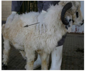 | |
| Fig. 1: | Staring and draggy hair coat and local hyperhidrosis (arrow) in affected ram |
RESULTS AND DISCUSSION
Laboratory examinations of skin scrapings confirmed infestation with Psoroptes ovis. Histopathologic findings included dilation of sweat glands (Fig. 2), hyperplasia of sebaceous glands, hyperkeratosis, ulcer and scab formation (Fig. 3) and eosinophilic dermatitis (Fig. 4). History and clinical findings association with results of
skin scraping and histopathologic findings indicated localized seborrhoeic dermatitis with hyperhidrosis. After treatment with ivermectin at dose rate of 0.2 mg kg-1, all clinical signs subsided. This confirmed that the cause of seborrhoeic dermatitis and hyperhidrosis was mite infestation and other possible causes were ruled out. Psoroptic mange is of greatest importance in sheep, in which it causes sheep scab, but it is also responsible for body mange in cattle and horses and ear mange in horses, sheep, goats and rabbits. Psoroptic mange is a major disease in sheep which was once virtually eliminated in most developed countries where wool production is an important industry. With the cessation of organophosphate dips in some countries there has been a resurgence of the problem (Radostits et al., 2007). Psoroptic mites abrade the surface and feed on lipid exudate, bacteria and skin debris. They cause the formation of scabs, under which they live. The eggs are laid on the skin at the edge of a scab and hatch in 1-3 days, although this is prolonged if eggs are not in contact with the skin (Guillot, 1981). The mite migrates to all parts of the skin and prefers areas covered with hair or wool.
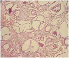 | |
| Fig. 2: | Dilatation of sweet glands, H&E x100 |
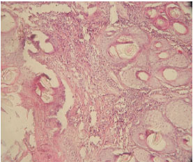 | |
| Fig. 3: | Hyperplasia of sebaceous glands, hyperkeatosis, ulcer and scab formation, H&E x100 |
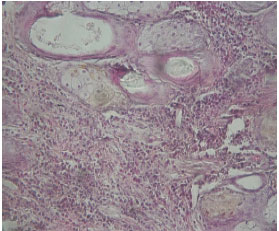 | |
| Fig. 4: | Eosinophilic dermatitis, H&E x100 |
Salivary secretions and mite excreta contain proteinases that result in a severe allergic pruritis. In allergic pruritus releasing of histamine can lead to increasing of vascular permeability (Scott, 1988). These can result dermatitis and hyperhidrosis. The exudation of serum accumulates to form a crust. In sheep the mites are more generally distributed and bacterial invasions of the skin are more common (Losson et al., 1999). Cutaneous lesions may occur on any part of the body but characteristically in badly affected sheep they are most obvious on the sides. Attention may be attracted to the area by raggedness of the wool caused by biting and scratching. In older lesions thin yellow crusts are present and the wool commences to shed. The wool may contain large masses of scab material which bind the fibers together in a mat. Behavioral changes in infested sheep are dramatic with sheep biting at the affected areas, rubbing or scratching. In addition infested sheep exhibited stereotypic behaviors typical of animals under stress. The mites can be easily demonstrated in scrapings taken from the edges of the lesions. Examination is facilitated by prior digestion of the scraping in warm, 10% potassium hydroxide solution (Falconi et al., 2002). Seborrhoeic dermatitis in a goat due to Malassezia pachydermatis has been reported by Pin (2004). Seborrheic dermatitis in a pygmy goat due to Staphylococcus hyicus has been recorded by Schamber and Alstad (1989). Localized seborrhoeic dermatitis association with hyperhidrosis due to mite infestations has never been reported in animals. So, this is the first report of localized seborrhoeic dermatitis association with hyperhidrosis due to mite infestation in animals.
REFERENCES
- Falconi, F., H. Ochs and P. Deplazes, 2002. Serological cross-sectional survey of psoroptic sheep scab in Switzerland. Vet. Parasitol., 109: 119-127.
PubMedDirect Link - Guillot, F.S., 1981. Population increase of Psoroptes ovis (Acari: Psoroptidae) on stanchioned cattle during summer. J. Med. Entomol., 18: 44-47.
PubMed - Losson, B.J., J.F. Lonneux and M. Lekimme, 1999. The pathology of Psoroptes ovis infestation in cattle with a special emphasis on breed difference. Vet. Parasitol., 83: 219-229.
PubMedDirect Link - Pin, D., 2004. Seborrhoeic dermatitis in a goat due to Malassezia pachydermatis. Vet. Dermatol., 15: 53-56.
PubMedDirect Link - Schamber, G. and A.D. Alstad, 1989. Isolation of Staphylococcus hyicus from seborrheic dermatitis in a pygmy goat. J. Vet. Diag. Invest., 1: 276-276.
PubMedDirect Link