
S. Sulochana
P.G. and Research Department of Zoology, Rajah Serfoji Government College Thanjavur, 6143001, Tamil Nadu, India
Palaniyandi Krishnamoorthy
P.G. and Research Department of Zoology, Rajah Serfoji Government College Thanjavur, 6143001, Tamil Nadu, India
K. Sivaranjani
P.G. and Research Department of Biotechnology, Nehru Memorial College, Puthanampatti, Tiruchirappalli-7, Tamil Nadu, India
Journal of Pharmacology and Toxicology
Year: 2012 | Volume: 7 | Issue: 5 | Page No.: 251-258


ABSTRACT
In the present investigation, an attempt was made to prepare nanoparticle by using a medicinally important plant Andrographis paniculata. Because now the biologically synthesized nanoparticle have been widely used in the field of medicine. Silver nitrate (AgNO3) was used to synthesis the silver nanoparticle by using leaf extract of Andrographis paniculata. The synthesized silver nanoparticle from 1 mM AgNO3 solution through the leaf extract were characterized using UV-Vis Spectrophotometry, XRD, SEM and FTIR. X-ray diffraction and SEM analysis showed the average particle size of 28 nm with cubic and hexagonal shape and it confirmed the formation of nanoparticle in the sample. The synthesized silver nanoparticle can be used for various applications due to its eco-friendliness, non toxic and compatibility for pharmaceutical and other applications.
PDF Abstract XML References Citation
Received: February 02, 2012;
Accepted: April 06, 2012;
Published: June 26, 2012
How to cite this article
S. Sulochana, Palaniyandi Krishnamoorthy and K. Sivaranjani, 2012. Synthesis of Silver Nanoparticles using Leaf Extract of Andrographis paniculata. Journal of Pharmacology and Toxicology, 7: 251-258.
DOI: 10.3923/jpt.2012.251.258
URL: https://scialert.net/abstract/?doi=jpt.2012.251.258
DOI: 10.3923/jpt.2012.251.258
URL: https://scialert.net/abstract/?doi=jpt.2012.251.258
INTRODUCTION
There is growing need to develop eco-friendly and body benign nanoparticle synthesis process without use of toxic chemicals in the synthesis of protocols to avoid adverse effects in biomedical applications (Ankamwar, 2010). Nanometal particles, especially silver, have drawn the attention of researchers (Safaepour et al., 2009). Because of their extensive application in the development of new technologies in the areas of electronics, material sciences and medicine at the nanoscale (Magudapathy et al., 2001). Silver nanoparticles have many applications; for example, they might be used as spectrally selective coatings for solar energy absorption and intercalation material for electrical batteries, as optical receptors, as catalysts in chemical reactions, for biolabelling and as antimicrobials (Joerger et al., 2000; Panacek et al., 2006). Many reports well documented on the biogenesis of silver nanoparticles using several plant extracts. The reducing property of different plant constituents may play a critical role in the reduction of Ag+ to silver nanoparticles (Shankar et al., 2004). The use of environmentally benign materials like plant leaf extract (Parashar et al., 2009), bacteria (Saifuddin et al., 2009), fungi (Bhainsa and D’Souza, 2006) and enzymes (Willner et al., 2007) for the synthesis of silver nanoparticles offers numerous benefits of eco-friendliness and compatibility for pharmaceutical and other biomedical applications as they do not use toxic chemicals for the synthesis protocol. Chemical synthesis methods lead to presence of some toxic chemical absorbed on the surface that may have adverse effect in the medical applications. Green synthesis provides advancement over chemical and physical method as it is cost effective, environment friendly, easily scaled up for large scale synthesis and in this method there is no need to use high pressure, energy, temperature and toxic chemicals. Therefore, the objective of this present study was to synthesis the biologically active nanoparticle from the leaf extract of medicinal plant Andrographis paniculata.
MATERIAL AND METHODS
Plant materials: The plant Andrographis paniculata were collected from the campus of Rajah Serfoji Govt. College in Thanjavur (DT) Tamil Nadu during October to December 2011.
Preparation of the extract: Andrographis paniculata leaf were collected and used to prepare the aqueous extract. Leaf weighing 25 g were thoroughly washed in distilled water, dried, cut into fine pieces and were crushed into 100 mL sterile distilled water and filtered through Whatman No.1 filter paper (pore size 25 μm). The filtrate was further filtered through 0.6 μm sized filters.
Synthesis of silver nanoparticles: One millimole aqueous solution of silver nitrate (AgNO3) was prepared and aqueous extract of leaf of Andrographis paniculata used for the synthesis of silver nanoparticles. 10 mL of Andrographis paniculata leaf extract was added into 90 mL of aqueous solution of 1 mM silver nitrate for reduction into Ag+ ions and kept at room temperature for 5 h.
UV-Vis spectra analysis: The reduction of pure Ag+ ions was monitored and measured in the UV-Vis spectrophotometer UV-2450 (Shimadzu).
XRD measurement: The silver nanoparticle solution thus obtained was purified by repeated centrifugation at 5000 rpm for 20 min followed by redispersion of the pellet of silver nanoparticles into 10 mL of deionized water. After freeze drying of the purified silver particles, the structure and composition were analyzed by XRD and SEM. The dried mixture of silver nanoparticles was collected for the determination of size of Ag nanoparticles. Pro X-ray diffract meter operated at a voltage of 40 kV and a current of 30 mA with Cu Kα radiation in a θ-2θ configuration. The crystallite domain size was calculated from the width of the XRD peaks, assuming that they are free from non-uniform strains, using the Scherrer formula:
where, D is the average crystallite domain size perpendicular to the reflecting planes, λ is the X-ray wavelength, β is the Full Width at Half Maximum (FWHM) and θ is the diffraction angle.
Scanning electron microscopic (SEM) analysis of silver nanoparticles: SEM analysis was done using Hitachi S-4500 SEM machine. Thin films of the sample were prepared on a carbon coated copper grid by just dropping a very small amount of the sample on the grid, extra solution was removed using a blotting paper and then the film on the SEM grid were allowed to dry by putting it under a mercury lamp for 5 min.
FTIR analysis of dried biomass after bioreduction: The residual solution of 100 mL after reaction was centrifuged at 5000 rpm for 10 min and the resulting suspension was redispersed in 10 mL sterile distilled water. The centrifugation and redispersing process was repeated three times. Thereafter, the purified suspension was freeze dried to obtain dried powder. Finally, the dried nanoparticles were analyzed by FTIR, Nicolet Avatar 660 (CECRI, Karaikudi).
RESULTS
The synthesized nanoparticles using Andrographis paniculata leaf extract exhibited a reddish brown color in aqueous solution due to excitation of surface plasmon vibrations. While the leaf extract of Andrographis paniculata mixing with the aqueous solution of the silver ion complex, it started to change the color from watery to yellowish brown due to reduction of silver ion (Fig. 1). The absorption spectrum of aqueous silver nitrate only solution exhibited maximum at about 220 nm and there was no color change. The synthesized AgNO3 nanoparticle were detected by UV-Vis spectroscopy at various nm 200, 300, 400, 500 and 600 nm, the particle has increasingly sharp absorbance maximum peak at 280 nm and gradually decreased while nanometer increased (Fig. 2). Andrographis paniculata was found to exhibit very strong absorption peaks at 400 to 500 nm. Absorption spectra of Ag nanoparticles formed on the reaction mixture at different time intervals at 280 nm. The particle has increasing sharp between 1 to 5 h the gradually decreased while the time has increased. The UV-Vis spectra recorded from the reaction medium after 5 h, the light absorption was more due to presence of nanoparticle formed in the reaction media and were showed strong absorbance peaks at 280 nm (Fig. 2). Broadening of peak indicated that the particles are poly dispersed. The biosynthesized silver nanostructure by employing Andrographis paniculata leaf extract was further demonstrated and confirmed by the characteristic peaks observed in the XRD image (Fig. 3) and the structural view were observed under the scanning electron microscope (Fig. 4). The XRD pattern showed three intense peaks in the whole spectrum of 2θ values ranging from 10 to 80. The typical XRD pattern (Fig. 3) reveled that the sample contains a mixed phase (cubic and hexagonal) structures of silver nanoparticles (Fig. 4). The average estimated particle size of this sample was 28 nm derived from the FWHM of peak (Fig. 3). The SEM image showed the high density silver nanoparticles synthesized by the A. paniculata development of silver nanostructures. FTIR analysis was used for the characterization of nanoparticles.
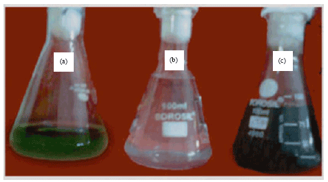 | |
| Fig. 1: | Synthesis of nanoparticle using silver nitrate and leaf extract of Andrographis paniculata a: Leaf extract, b: 1 mM AgNO3 without leaf extract and c: 1 mM AgNO3 with 10% leaf extract after 5 h |
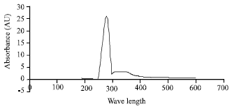 | |
| Fig. 2: | UV-Vis absorption spectrum of leaf extract synthesized by treating 1 mM aqueous AgNO3 solution with 10% Andrographis paniculata leaf extract after 5 h |
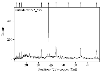 | |
| Fig. 3: | XRD pattern of silver nanoparticles synthesized by treating 10% Andrographis paniculata with 1 mM aqueous AgNO3 solution |
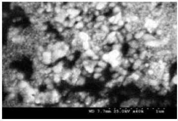 | |
| Fig. 4: | SEM micrograph of silver nanoparticles |
FTIR absorption spectra of water soluble extract before and after reduction of Ag ions are shown in Fig. 5. Absorbance bands in Fig. 5a (before bioreduction) are observed in the region of 500-3500 cm-1 are 3354.04, 2925.22, 2858.42, 1650.24, 1541.04, 1450.97, 1323.46, 1244.31, 1153.67, 1030.04, 608.35 cm-1.
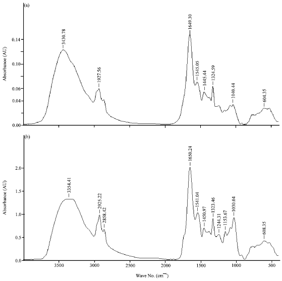 | |
| Fig. 5(a-b): | FTIR graph of (a) Leaf extract and (b) Nanoparticles synthesized in 10% leaf extract solution |
These absorbance bands are known to be associated with the stretching vibrations for -C C-C O, -C C- [(in-ring) aromatic], -C-C- [(in-ring) aromatic], C-O (esters, ethers) and C-O (polyols), respectively. In particular, the 1244 cm-1 band arises most probably from the C-O group of polyols such as hydroxyflavones and catechins. The total disappearance of this band after the bioreduction (Fig. 5b).
DISCUSSION
Silver nanoparticle was synthesized from the leaf of Andrographis paniculata. Rao and Savithramma (2011) revealed the synthesis nanoparticles from leaf of Svensonia hyderabadensis and leaves of Allium cepa by Saxena et al. (2010) and Clerodendrum inerme by Farooqui et al. (2010) and in Argemone mexicana (Khandelwal et al., 2010). Leaf extract of Andrographis paniculata while mixing with the aqueous solution of the silver ion complex, it started to change the color from watery to yellowish brown. Which indicated the formation of silver nanoparticles and this might be due to the reduction of silver ion to form the nanoparticles. Similarly, Elumalai et al. (2010) observed that the reduction of silver ion in Ag nitrate exposed plant extract followed by color change and Rao and Savithramma (2011) also reported that the Svensonia hyderabadensis solution of the silver ion complex started to change the colour from yellow to dark brown due to reduction of silver ions. Silver nanoparticles exhibited yellowish brown colour in aqueous solution due to excitation of surface plasmon vibrations (Shankar et al., 2004). Chen et al. (2002) observed the intensity of color development in the reaction mixture of different plants such as in Helianthus annuus (sunflower), Basella alba (spinach) and Saccharum officinarum (sugarcane). Further, who stated that the synthesized AgNO3 nanoparticle were detected by UV-Vis spectroscopy at various nanometers and the particle has increasingly sharp absorbance and the peak was maximum at 280 nm and then it was gradually decreased while nanometer increased. Andrographis paniculata was also found to exhibit very strong absorption peaks at 400 to 500 nm. and the absorption spectrum of aqueous silver nitrate only solution exhibited maximum at about 220 nm and there was no color change. Rao and Savithramma (2011) reported that the UV-Vis spectrophotometric measurements were showed strong absorption peak at 300 to 400 nm.
Chen et al. (2002) suggested that the difference in the morphology of the synthesized nanoparticle might be a reason for the variations in the absorption peaks.
The biosynthesized silver nanostructure by employing Andrographis paniculata leaf extract was further demonstrated and confirmed by the characteristic peaks observed in the XRD image and the structural view were observed under the scanning electron microscope. The average estimated particle size of this sample was 28 nm derived from the FWHM of peak. The SEM image showed the high density silver nanoparticles synthesized by the A. paniculata development of silver nanostructures. FTIR analysis was used for the characterization of the extract and the resulting nanoparticles. FTIR absorption spectra of water soluble extract before and After reduction of Ag ions. Absorbance bands are observed in the region of 500-3500 cm-1. Saifuddin et al. (2009) observed the FTIR measurements to identify the possible biomolecules responsible for capping and efficient stabilization of the metal nanoparticles synthesized in leaf broth. The beaks near 3354.04, 2925.22 and 2858.42 cm-1 assigned to OH stretching and aldehydic C-H stretching, respectively (Jain et al., 2009). The total disappearance of this band after the bioreduction may be due to the fact that the polyols are mainly responsible for the reduction of Ag ions, where by they themselves get oxidized to unsaturated carbonyl groups leading to a broad peak at 1650.24 cm-1. The weaker band at 1244.31 cm-1 corresponds to amide I arising due to carbonyl stretch in proteins (Sathyavathi et al., 2010). The result of all earlier research strongly support the need of preparation of silver nanoparticle from the medicinally important medicinal plants for various medicinal uses.
CONCLUSION
Synthesis of silver nanoparticle from the leaf of Andrographis paniculata was confirmed by the colour changes from yellow to dark brown. Which indicated the formation of silver nanoparticles. Therefore, the growing need of developing a eco-friendly nanoparticle synthesis is possible and it can be used for various biomedical applications to avoided the adverse effects chemically synthesized nanoparticle in the filed of medical applications.
ACKNOWLEDGMENTS
The authors are thankful to the U.G.C, New Delhi for providing financial assistance and the Director, Central Electro Chemical Research Institute, Karaikudi, Tamil Nadu for providing analytical facilities.
REFERENCES
- Ankamwar, B., 2010. Biosynthesis of gold nanoparticles (green-gold) using leaf extract of Terminalia catappa. E-J. Chem., 7: 1334-1339.
Direct Link - Bhainsa, K.C. and S.F. D'Souza, 2006. Extracellular biosynthesis of silver nanoparticles using the fungus Aspergillus fumigatus. Colloids Surf. B: Biointerfaces, 47: 160-164.
CrossRefDirect Link - Elumalai, E.K., T.N.V.K.V. Prasad, J. Hemachandran, S.V. Therasa, T. Thirumalai and E. David, 2010. Extracellular synthesis of silver nanoparticles using leaves of Euphorbia hirta and their antibacterial activities. J. Pharm. Sci. Res., 2: 549-554.
Direct Link - Farooqui, M.A., P.S. Chauhan, P. Krishnamoorthy and J. Shaik, 2010. Extraction of silver nanoparticles from the left extracts of Clerodendrum incerme. Digest J. Nanomater. Biostruct., 5: 43-49.
Direct Link - Singh, A., D. Jain, M.K. Upadhyay, N. Khandelwal and H.N. Verma, 2010. Green synthesis of silver nanoparticles using Argemone Mexicana leaf extract and evaluation of their antimicrobial activities. Digest J. Nanomater. Biostruct., 5: 483-489.
Direct Link - Saxena, A., R.M. Tripathi and R.P. Singh, 2010. Biological synthesis of silver nanoparticles by using onion (Allium cepa) extract and their antibacterial activity. Digest J. Nanomater. Biostruct., 5: 427-432.
Direct Link - Joerger, R., T. Klaus and C.G. Granqvist, 2000. Biologically produced silver-carbon composite materials for optically functional thin-film coatings. Adv. Mater., 12: 407-409.
CrossRefDirect Link - Magudapathy, P., P. Gangopadhyay, B.K. Panigrahi, K.G.M. Nair and S. Dhara, 2001. Electrical transport studies of Ag nanoclusters embedded in glass matrix. Phys. B: Condens. Matter, 299: 142-146.
CrossRefDirect Link - Safaepour, M., A.R. Shahverdi, H.R. Shahverdi, M.R. Khorramizadeh and A.R. Gohari, 2009. Green synthesis of small silver nanoparticles using geraniol and its cytotoxicity against Fibrosarcoma-Wehi 16. Avicenna J. Med. Biotechnol., 1: 111-115.
Direct Link - Panacek, A., L. Kvitek, R. Prucek, M. Kolar and R. Vecerova et al., 2006. Silver colloid nanoparticles: Synthesis, characterization and their antibacterial activity. J. Phys. Chem. B, 110: 16248-16253.
CrossRefPubMedDirect Link - Saifuddin, N., C.W. Wong and A.A.N. Yasumira, 2009. Rapid biosynthesis of silver nanoparticles using culture supernatant of bacteria with microwave irradiation. J. Chem., 6: 61-70.
CrossRefDirect Link - Sathyavathi, R., M.B. Krishna, S.V. Rao, R. Saritha and D.N. Rao, 2010. Biosynthesis of silver nanoparticles using coriandrum sativum leaf extract and their application in nonlinear optics. Adv. Sci. Lett., 3: 138-143.
CrossRefDirect Link - Shankar, S.S., A. Rai, A. Ahmad and M. Sastry, 2004. Rapid synthesis of Au, Ag, and bimetallic Au core-Ag shell nanoparticles using Neem (Azadirachta indica) leaf broth. J. Colloid Interface Sci., 275: 496-502.
CrossRefDirect Link - Willner, B., B. Basnar and B. Willner, 2007. Nanoparticle-enzyme hybrid systems for nanobiotechnology. FEBS J., 274: 302-309.
CrossRefDirect Link - Chen, S., S. Webster, R. Czerw, J. Xu and D.L. Carroll, 2002. Morphology effects on the optical properties of silver nanoparticles. J. Nanosci. Nanotechnol., 4: 254-259.
PubMedDirect Link