
Jahanshahi Mehrdad
Department of Anatomy, Gorgan University of Medical Sciences, Gorgan, Iran
Y. Sadeghi
Department of Anatomy, Shahid Beheshti University of Medical Sciences, Tehran, Iran
Journal of Medical Sciences
Year: 2007 | Volume: 7 | Issue: 3 | Page No.: 457-459


ABSTRACT
The subclavian artery is a major branch of the Aorta. In the left, the subclavian artery arises from Aortic arch and in the right it arises from the Brachiocephalic trunk. This artery passes from the subclavian groove on the first rib and continues as Axillary artery. The subclavian artery has several branches that supply chest wall, Thyroid gland and cervical regions. Several variations about the subclavian artery and it`s branches were have been reported. In this case, from the first part of this artery, we found a common trunk that gives the ascending cervical, Transvers cervical, Suprascapular and Dorsal scapular arteries. The inferioe thyroid artery was absent.
PDF Abstract XML References Citation
How to cite this article
Jahanshahi Mehrdad and Y. Sadeghi, 2007. A Case Report of Variation in Subclavian Artery Branches. Journal of Medical Sciences, 7: 457-459.
DOI: 10.3923/jms.2007.457.459
URL: https://scialert.net/abstract/?doi=jms.2007.457.459
DOI: 10.3923/jms.2007.457.459
URL: https://scialert.net/abstract/?doi=jms.2007.457.459
INTRODUCTION
The subclavian and axillary arteries are gaining increasing interest in the cardiac surgery community as they have been successfully used as cannulation sites for cardiopulmonary bypass in thoracic aortic procedures and redo operations. They have also been taken for insertion of intraaortic balloon pumps and most recently they are under discussion for use as an inflow vessel in coronary artery surgery (Bonatti and Coulson, 2000).
The subclavian artery, in the left arises from Aorta and in the right, behind of the right sternoclavicular joint, arises from braciocephalic trunk. This artery passes from the superior surface and outer border of the first rib and continue as axillary artery.
The subclavian artery, for purposes of description, is divided into three parts by the scalenus anterior muscle, which crosses the artery anteriorly. The first part of the artery extends from its origin to the medial border of the scalenus anterior. The second part lies posterior to this muscles and the third part extends from the lateral border of the scalenus anterior to the outer border of the first rib (Williams et al., 1995).
The branches of the subclavian artery are as follows:
First part: Vertebral artery, Internal thoracic and Thyrocervical trunk that gives off three branches: Inferior thyroid artery, Superficial cervical and Suprascapular arteries.
Second part: The Costocervical trunk, that it divides into the superior intercostals and Deep cervical arteries.
Third part: Has no branches and occasionally the Superficial cervical or the suprascapular artery, or both, arise from this part (Williams et al., 1995).
Case report: During routin dissection for undergraduate students in dissection room of Shahid Beheshti University of medical Sciences in Tehran, Iran in one old man cadaver, we found that the pattern of branches in the left Subclavian artery is abnormal.
In this case almost, all of the branches of the subclavian arteries arises from the first part of it. Vertebral and Internal thoracic arteries are normally but a big common trunk arises from first part, that it is similar to thyrocervical trunk in normally, but any branches of its don't go to the thyroid gland, in the other hand, the inferior thyroid artery is absence. Then this trunk divides to Ascending cervical artery and transvers cervical artery.
Then the transvers cervical artery goes to the posterior triangle of the neck and divides to suprascapular and dorsal scapular arteries.
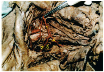 | |
| Fig. 1: | Left subclavian branches in our case, the arteries showed by red, 1 = Common trunk, 2 = Suprascapular artery, 3 = Dorsal scapular and 4 = Muscular branch |
As the second and third parts of subclavian artery in this case have not any branches, the costocervical trunk arises from the dorsal scapular artery (Fig. 1).
DISCUSSION
Variation in branches of subclavian artery have been reported (Bergman and Ryosuke, 2002a, b). for example in one case, the right subclavian artery as the last branch of Aorta, was retropharyngeal and this position exist in less than one percent (Almenar-Garcia et al., 2002; Komiyama et al., 1995).
In some cases the internal thoracic artery arises from the third part of subclavian artery and the left vertebral artery arises correctly from the Aortic arch between the left common carotid and the left subclavian artery (Vorster et al., 1998).
Occationally, the thyrocervical trunk is absent and it's branches arise from the other arteries, for example the transvers cervical artery arise from the subclavian and the suprascapular artery arises from the internal thoracic artery (Yucel et al., 1999).
In our case, a special variation is reported that it is differ from the other cases that they reported previously.
REFERENCES
- Bonatti, J. and A.S. Coulson, 2000. The subclavian and axillary arteries as inflow vessels for coronary artery bypass grafts combined experience from three cardiac surgery centers. Heart Surg. Forum, 3: 307-312.
Direct Link - Komiyama, M., Y. Matsuno and Y. Shimada, 1995. Variation of the right subclavian artery as the last branch of the aortic arch in two Japanese cadavers. Okajimas Folia Anat Jpn., 72: 245-247.
PubMedDirect Link - Vorster, W., P.T. du Ploy and J.H. Mreiring, 1998. Abnormal origin of internal thoracic and vertebral arteries. Clin Anat, 11: 33-37.
CrossRefDirect Link - Yucel, A.H., E. Kizilkanat and C.O. Ozdemir, 1999. The variation of the subclavian artery and it's branches. Okajimas Folia Anat Jpn., 76: 255-261.
Direct Link