
W. Aengwanich
Stress and Oxidative Stress Research Unit, Faculty of Veterinary Medicine and Animal Science, Mahasarakham University, Maha Sarakham 44000, Thailand
N. Narkkong
Central Instrumentation Unit, Faculty of Science, Mahasarakham University, Maha Sarakham 44150, Thailand
A. Tanomthong
Department of Biology, Faculty of Science, Khonkhaen University, Khon Khaen 40002, Thailand
International Journal of Zoological Research
Year: 2008 | Volume: 4 | Issue: 1 | Page No.: 68-71


ABSTRACT
Blood samples from eastern sarus cranes housed at Nakhonratchasima Zoo, Nakhon Ratchasima, northeastern, Thailand were collected in January, 2007. A morphological observation of the thrombocytes was examined using scanning electron microscopy. The results revealed the following information: The thrombocytes of the eastern sarus cranes are tiny cells and they display a smooth, irregular, spherical and pseudopodic membrane surface with long microfilament protrusions from their membrane. The thrombocytes of the eastern sarus cranes use these microfilaments to attach to other blood cells for movement and blood clumping. The long microfilaments were not found in fish, reptiles and other domestic birds. The morphological structure of the thrombocyte, the microfilaments and the activities of the eastern sarus crane has not been reported previously.
PDF Abstract XML References Citation
How to cite this article
W. Aengwanich, N. Narkkong and A. Tanomthong, 2008. Morphological Observations on the Thrombocyte of Eastern Sarus Cranes (Grus antigone sharpii) in Northeastern Thailand. International Journal of Zoological Research, 4: 68-71.
DOI: 10.3923/ijzr.2008.68.71
URL: https://scialert.net/abstract/?doi=ijzr.2008.68.71
DOI: 10.3923/ijzr.2008.68.71
URL: https://scialert.net/abstract/?doi=ijzr.2008.68.71
INTRODUCTION
The eastern sarus crane (Grus antigone sharpii) is the world`s tallest flying bird (Jeltes, 2006). As one of three Grus antigone subspecies, it formerly inhabited Indochina; however, it`s population there has been greatly reduced in distribution and number. Flocks have been recently located in Myanmar, Cambodia and Vietnam, but it appears as if the population may now be fragmented into two separate sub-populations. Total numbers are estimated to be between 1,000 and 1,600 birds (Zoo, 1997). Jeltes (2006) reported that in this decade the total population of the eastern sarus crane has been reduced to between 500-1,500 birds. At present, sarus cranes are currently listed as vulnerable A1cde + 2cde on the IUCN red list. Nakhonratchasima Zoo obtained their eastern sarus cranes from the forests in Cambodia and keeps them in cages in the breeding section and public viewing section of the Zoo. At present, Nakhonratchasima Zoo has more than 40 eastern sarus cranes.
Thrombocytes have their origins in bone marrow and play a primary role in hemostasis (Sturkie, 1965) clot promotion (Shepro et al., 1969) and tendency to clump in peripheral blood films and may also have a phagocytic function to aid in the removal of foreign material from the blood (Campbell, 1995). Whereas, documents about the thrombocyte`s characteristics in the eastern sarus crane are limited, the objective of this preliminary study was to examine the morphological aspect of the thrombocytes in eastern sarus crane by using SEM. Basic knowledge from this examination is important to any future in depth study of the hematology of this bird.
MATERIALS AND METHODS
Birds
Two eastern sarus cranes (male and female) were reared in the breeding section of Nakhonratchasima Zoo, Nakhon Ratchasima Province northeastern Thailand. Their blood samples were collected in January, 2007. Cranes were restrained manually and five milliliters of blood sample were collected from the wing vein (Rithchie et al., 1994) using a 10 mL syringe, 21 gauge needle and 1.5 inch of length then placed in plastic tube with EDTA. The samples were cooled to approximately 4 °C (Campbell, 1995) using icepacks and then taken to the Central Instrumentation Units, Faculty of Science, Mahasarakham University within 12 h of blood collection.
SEM Technique
Blood samples were dropped in 2.5% glataraldehyde in 0.1 M phosphate buffer, pH 7.2 overnight at 4 °C then washed in the same buffer. Samples were postfixed with 1% Osmium tetroxide for 2 h then rinsed with distilled water, dehydrated in 20, 40, 60, 80 and 100% acetone and left to air dry. Gold coated blood films were examined with a SEM (JSM 6460LV).
RESULTS AND DISCUSSION
Thrombocytes of the eastern sarus crane were examined by using SEM. The results revealed the following information: The thrombocytes are tiny cells (diameter = 2-3 μm); with a smooth, irregular and pseudopodic membrane surface with long microfilaments protruding from the membrane (Fig. 1A-H). The cell size of the thrombocytes in the eastern sarus crane were smaller than that of the chickens by about 2 times. Daimon and Uchida (1978) found that mature thrombocytes of the white leghorn chickens measured approximately 10 μm in length and 5 μm in width. Salakij et al. (2002) reported that the thrombocytes of the king cobra had an elongated shape with a smooth membrane. Moreover, Santos et al. (2003) and Sano-Martins et al. (1994) found that the thrombocytes of snakes and the roadside hawk, normally ellipitical in shape, become spheroid with cytoplasmic protrusions and adhere to one another. This was accordant with the shape of the thrombocytes of the eastern sarus cranes in this study.
The membrane surface of the thrombocyte of the eastern sarus crane was similar to that reported by Shepro et al. (1969) and Maxwell (1974). They found that under ultra-structural observation, the membrane surface in dogfish and domestic birds had many forms: smooth, irregular, pseudopodia, lobopodia and villi like; whereas, the long microfilaments, which were found in the eastern sarus cranes, were not found in fish, reptiles and other domestic birds. Figure 1 showed that the thrombocytes of the eastern sarus cranes used their microfilaments to attach to other blood cells. Moreover, the thrombocytes used these structures for blood clumping.
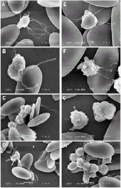 | |
| Fig. 1: | SEM micrograph of thrombocyte in eastern sarus cranes (Grus antigone sharpie) (A) normal thrombocyte with long microfilament, (B) thrombocyte with microfilament attached to leukocyte and red blood cell, (C) clumping of thrombocyte, (D) thrombocyte with microfilament attached to mature and young red blood cell, (E) thrombocyte with microfilament attached to red blood cell, (F) thrombocyte with microfilament attached to red blood cell, (G) thrombocte with microfilament and (H) clumping of thrombocyte (E, F and G = 1 μm; A, B and H = 2 μm; C and D =5 μm) |
CONCLUSIONS
The thrombocytes of the eastern sarus cranes are small and their surface membrane had several forms with long microfilament protrusions from their membrane. They used the microfilaments for blood clumping and to attach to other blood cells. No previous reports on the morphological structure of the thrombocytes, the microfilaments or the activities of the eastern sarus cranes have been published.
REFERENCES
- Daimon, T. and K. Uchida, 1978. Electron microscopic and cytochemical observations on the membrane systems of the chicken thrombocyte. J. Anat., 125: 11-21.
Direct Link - Maxwell, M.H., 1974. An ultrastructural comparison of the mononuclear leucocytes and thrombocytes in six species of domestic bird. J. Anat., 117: 69-80.
Direct Link - Salakij, C., J. Salakij, S. Apibal, N. Narkkong, L. Chanhome and N. Rochanapat, 2002. Hematology, morphology, cytochemical staining, ultrastructural characteristics of blood cell in king cobras (Ophiophagus hannah). Vet. Clin. Pathol., 31: 116-126.
CrossRef - Sano-Martins, I.S., C. Jared and A. Jr. Brunner, 1994. Ultrastructural alterations in thrombocytes of the snake Waglerophis merremii after activation by ADP. Comp. Haematol. Int., 4: 226-231.
CrossRefDirect Link - Santos, A.A., A.M. Joppert da Silva, V.A. Lima de Carvalho and M.I. Egami, 2003. Morpho-cytochemical and ultrastructural characterization of peripheral thrombocytes of roadside hawk (Buteo margnirostris). Int. J. Morphol., 21: 279-284.
Direct Link