
Sarma Nursani Lumbanraja
Department of Obstetrics and Gynecology, University of Sumatera Utara, Medan, Indonesia
International Journal of Osteoporosis and Metabolic Disorders
Year: 2016 | Volume: 9 | Issue: 1 | Page No.: 13-18


ABSTRACT
Background: Steroid had become the most consumed drug that can be obtained easily. However, its wide range of side effects is not known by its user. Bone loss is one of the side effects that can cause debilitation either in physic or psychical term of a person. Objective: To determine the difference of steroid used between osteoporosis and control group. Methodology: This was an analytical, case control study that conducted at South Securai Village, Langkat, Indonesia from February-March 2016. This study included each 21 osteoporosis and normal reproductive women. The questionnaires were given to all subjects, to assess demographic data, knowledge, behaviour, steroid usage and factors influenced the use of steroid. Bone mass density was measured by a standardized scale. Data was processed by statistical product and service solutions (SPSS) 22.0 with 95% CI and p<0.05 was considered significant. Results: Baseline characteristics between the osteoporosis group and control group were simliar. This study showed significant difference of steroid used between osteoporosis and control group (66.7 vs 23.8%, p = 0.005). No differences of bone mass denisty was found regarding of how steroid being used. Conclusion: There was an association between steroid use and bone mass loss.
PDF Abstract XML References Citation
Received: May 25, 2016;
Accepted: June 03, 2016;
Published: June 15, 2016
How to cite this article
Sarma Nursani Lumbanraja, 2016. Effects of Steroid Use and Bone Mass Density in South Securai Village, Langkat, Indonesia. International Journal of Osteoporosis and Metabolic Disorders, 9: 13-18.
DOI: 10.3923/ijom.2016.13.18
URL: https://scialert.net/abstract/?doi=ijom.2016.13.18
DOI: 10.3923/ijom.2016.13.18
URL: https://scialert.net/abstract/?doi=ijom.2016.13.18
INTRODUCTION
Analgesic had become one of the top highly demand over-the-counter drugs in many countries, also in Indonesia1. Dale et al.2 reported prevalence of using over-the-counter analgesics at least once per week was 47%. Ely et al.3 showed that 28.8% of 758 elderly persons used 1-4 analgesics, which mostly were steroids. Although, OTCs are considered safe when used appropriately, they may cause serious multiorgan side effects3.
Steroids are used in almost all medical specialties. In US, reported 1.2% of the populations consume long term glucocorticoids4. In UK, Van Staa et al.5 reported percentages of 0.85-0.9%, mostly in aged 70-79 years people. In Northeast Iceland, the incidence6 was 0.7%. In the United Kingdom, it is showed that about 1% of the general population received steroid therapy in any point of time7.
Steroid had metabolic (glucocorticoid) and electrolyte-regulating (mineralocorticoid) activities8. Steroid began its action by binding to steroid receptors in the cytoplasm9. This marked the activation of steroid, which will then translocate to nucleus and form complex with Glucocorticoid Response Elements (GRE) in the promoter region of steroid-responsive genes10. The binding will lead to several effects. If the complex activated anti-inflammatory proteins secretory leukoprotease inhibitor and mitogen-activated protein kinase phosphatase-1 (MKP-1) which inhibits MAP kinase pathways, this will contribute to reduction of pain11. If the complex activated cis and trans-repression, NF-KB, POMC, CRF-1 and osteocalcin will reduce and cause antiinflammatory effect12.
Many side effects of steroids had been established because its wide range of action. Short term side effects included mild immunosupression, hyperglycemia and psychiatric disorders. Long-term side effects included peptic ulceration, osteoporosis and Cushing’s syndrome13. Van Staa et al.14 showed that using steroid can increase 1.33 risk of fracture. Osteoporosis can greatly increase the risk of fracture, which will profoundly impair daily activities, decrease the quality of life, dan life expectancy15. Studies of oral steroid dose and loss of bone mineral density have reported inconsistent results14.
Walsh et al.16 showed that steroid use cause lower bone mass density, ranged from 0.6-1.02 g cm‾2 in femoral neck. In contrast, Selby et al.17 found a similar frequency of vertebral fractures and bone mass in steroid user and control. Matsumoto et al.18 showed that this effect was due to type of steroid use. They showed bone mass loss in oral steroid user but not in inhaled steroid user, even in high dose. In other studies, bone mass was found to be loss only in specific location, mostly in spinal and femoral18.
Quantitative ultrasound of the os calcis is accepted as an effective low-cost method to assess osteoporotic fracture risk19. Based on ISCD recommendations, in situations where central DXA systems are not readily available, quantitative ultrasound can be used as an effective screening tool with >90% sensitivity for detecting patients with osteoporosis20.
MATERIALS AND METHODS
This was an analytical, case control study that conducted at South Securai Village, Langkat, Indonesia from February-March 2016. This study has been approved by the Ethical Committee of University of Sumatera Utara. In order to have a representative sample, minimal required samples were detemined and yielded 21 samples in each groups of study. This study was done simultaneously with the bone mass density check event that conducted in South Securai Village, Langkat, Indonesia. This study included 21 consecutive osteoporosis subjects and 21 controls. Women in reproductive age were chosen to ensure the pure effect to steroid in lower bone mass density, not the part of aging effect.
Subjects were all invited to participate in this study unless they had other reasons for fracture, for example, prolonged immobility, early menopause or taking of drugs that are known to affect BMD (anabolic steroids, sodium fluoride tablets, calcitonin, bisphosphonates (more than 1 month)), calcium supplements (more than 500 mg day–1 for more than 6 months), vitamin D (more than 400 IU day–1) or hormone replacement therapy (currently, more than 3 months in the past 10 years or more than 2 years ever).
A questionnaire containing demographic data (age, sex, educational level and job) and the use of medication was used for data collection. A questionnaire was developed to assess knowledge, behaviour and factors that influenced steroid usage. This questionnaires had been validated in this observational study before in 20 subjects. The questionnaire contained 5 questions for knowledge and 5 questions for behaviour (Table 1). In assessing knowledge of steroid side effects, subjects were being asked about steroid’s side effects such as hypertension, delayed growth, increase weight, moon face, osteoporosis and diabetes. If respondents know >3 of the side effects, we considered she know the side effects, vice versa. In determining influential factors, enabling factors (access to health care, payment of health care fee, availability of drug) and reinforcing factors (indication of joint pain, fatigue, increase stamina, increase sexua desire, willing to gain weight and increase of appettite) were analyzed. To assess steroid use, subjects were being asked the type of steroid they consumed, how routine the steroid be consumed and how long they have used steroid.
Bone mass density, weight and height was measured using Tabita Scale (Onemed), using the same scale for all patients. Daily calibration checks remained stable throughout the study. The clinical interpretation of osteoporosis was determined using the machine instructions. In female weight <50 kg, bone mass lower than 1.95, in female weight 50-70 kg, bone mass lower than 2.40, in female weight >70 kg, bone mass lower than 2.95 were considered osteoporosis.
Data were analyzed by SPSS (Statistical Product and Service Solutions, Chicago, IL, USA) 22.0 for Windows. Categorical data were expressed as number and continuous data as Mean±SD. Chi-square test (Fisher’s exact test) was used to examine the relation between qualitative variables. T-independent, Pearson correlation, Chi square, Fischer exact and ANOVA were used to evaluate quantitative variables. Significant value was take 95% and p<0.05 was considered significant.
RESULTS
Baseline characteristics between the osteoporosis group and control group were simliar (Table 2). It is found that a high steroids used in osteoporosis group instead of in NSAIDs group (66.7 vs 23.8%, p = 0.005).
All factors that influenced the NSAIDs and steroid usage in this study were assessed. Far access to primary care, health care fee, availability of steroid over the counter, were the factors that affected GC usage in most subjects (Table 3). The highest indication of GC usage were joint pain, fatigue and desire to increase stamina (all 94.7%, respectively).
The knowledge of steroid users in this study was in conflicting result with their behaviours. Majority of subjects showed bad knowledge, but they show a good behavior regarding of steroid usage (Table 1). If each of the knowledge questions were assessed separately, most subjects had lower knowledge in steroid’s side effects. Despite mostly know that steroid can cause weight gain (81%) and moon face (71.4%), all subjects (100%) do not know about the increase risk of diabetes mellitus in steroid usage. Rarely known side effects were delayed growth (4.8%), lower mass density (4.8%) and hypertension (33.3%). Majority also did not know type of steroid they consumed.
| Table 1: | Knowledge and behaviour of steroid in this study |
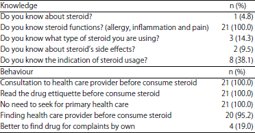 | |
| Table 2: | Baseline characteristics of this study |
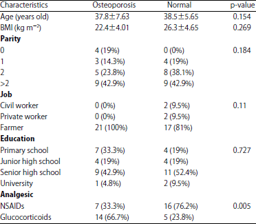 | |
| Table 3: | Factors that affected glucocorticoids usage |
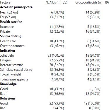 | |
| Table 4: | Association between steroid use and bone mass density |
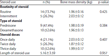 | |
To analyze the association between steroid use and bone mass density, statistical analysis was only conducted in osteoporosis group who used steroid. No differences of bone mass denisty was found regarding of type of steroid used (p = 0.384), steroid doses (p = 0.407), length of steroid use (p = 0.957) and routinity of steroid use (p = 0.425) (Table 4).
DISCUSSION
Steroids formed many compound, distributed in living creatures and based on the 1,2-cyclopentenophenanthrene skeleton21. As a drug, steroid can be classified as the short, intermediate and long acting. Short acting (<12 h) steroids include cortisol and cortisone with potency of 0.8-1. Intermediate acting steroids (12-36 h) include prednisolone, prednisone, methylprednisolone and triamcinolone with potency 0.8-6. Long acting steroids (>36 h) include dexamethasone and beclomethasone with potency9 of 0-25.
In an observational study priorly, it was found that almost all women in South Securai village consume analgesics. They suffered from joint and muscle pain due to their jobs as farmers. Indeed, steroids was largely used, in addition, they can obtain these drugs easily. In this study, majority of subjects (52.6%) use dexamethasone and the highest indication of sterois usage were joint pain, fatigue and desire to increase stamina. It seems that dexamethasone may be commonly used for pain management due to its high potency, long duration of action and minimal mineralocorticoid effect. Dexamethasone was shown to prolong axillary brachial plexus blockade if it was added to lidocaine22.
Side effects of steroids should be evaluated further of its highly usage. In this study, the knowledge regarding of the steroid’s side effects was low. This cause an extensive used of steroid among the subjects in this study. Steroids can cause emosional liability, insomnia, depression, glaucoma, peptic ulcer, fatty liver, visceral obesity, fluid retention, breakdown of protein, musclular atrophy, reduce bone formation, reduce bone mass, reduce linear growth and immunosuppression23.Study showed that side effects of using prednisolone and dexamethasone were similar, betamethasone showed greater side effects24.
Almost all subjects (95.2%) did not know about the effect of steroid inducing osteoporosis. Steroid-induced osteoporosis was discovered by Cushing more than 60 years ago25. Steroid had direct and indirect effects on osteoblasts, osteoclasts and osteocytes, leading to negative bone balance26. In osteocyte, steroid induce RANKL and M-CSF in osteocytes that will leads to apoptosis. In osteoblast, steroid induce activation of caspase 3, that witll reduce osteoblastogenesis, reduce bone formation and decrease bone qualities In osteoclast, steroid induce PPRAR-2, reduce WNT signal and decrease caspase 3 that will increase osteoclastogenesis. This will reduce bone mineral density and increase fracture risk. Steroid will also decrease bone resoprtion via neuroendocrine system. Besides that, it will decrease absorption of calcium from intestine and increase calcium excretion that will decrease bone calcium and increase PTH. Steroid will also cause proetolygiss of myofibrils and make risk of muscular weakness27. In men, glucocorticoids can inhibit testosterone production due to direct effect on the testis and indirect effects via suppression of gonadotropin hormone secretion. Low serum testosterone levels can contribute to the decrease of osteoblastic activation28.
Many studies had shown the higher effect of bone mass loss in steroid consumption. This study showed a significant association between glucocorticoid use and osteoporosis (p = 0.005). The steroid-induced bone loss loss is more pronounced in the first months of therapy, slowing down after about one year of therapy6. About 10-20% loss of trabecular bone occurs in the first 6 months of steroid use and 2% in subsequent. In addition, a 2-3% loss of cortical bone occurs in the 1st year, then a slow and continuous loss is maintained14,29. The earliest changes of steroid induced osteoporosis are usually seen in sites of high trabecular bone content such as the lumbar spine and ribs, but bone loss can occur at any site30.
Far access to primary care, health care fee, availability of steroid over the counter, were the factors that affected GC usage in most subjects. Although, behavior was considered good, most subjects admitted that they were better to find over the counter drugs for the complaints. This can be caused by far access to primary care (60.9%) and economics problem, as most subjects (84.2%) had to pay their health care fee by themselves.
Many studies found that risk of fracture depends on the steroid dose. Some studies showed that prednison 2.5 mg day–1 had 1.55, 2.5-7.5 mg day–1 had 2.59 and >7.5 mg day–1 had 5.18 risk of fracture. Three international guidelines had also made a cut off of daily 5 mg prednison consumption fort at least 3 months to receive further intervention in preventing osteoporosis. Other guidelines have recommended that prevention at doses31 ≥7.5 mg day–1. On the other hand, the UK Bone Research Society, the National Osteoporosis Society and the Royal College of Physician guidelines have not specified the dose32. Meta analysis by Van Staa et al.14 concluded that consumption of oral corticosteroid >5 mg day–1 leads to a reduction in bone mineral density and increase risk of fracture. Cumulative steroid dose was also correlated to the risk of fracture, but this correlation was weaker than that observed between daily dose and risk of fracture12. However, in this study, that the use of steroid, regardless of its doses (p = 0.407), length (p = 0.957) and daily (p = 0.425) use were all associated with decrease bone mass density. This can be caused by small sample size and the tendency of low bone mass density in the subjects in this study.
Although bone density testing is not recommended routinely for pre-menopausal women, it should be performed in women at risk for bone loss33. Densitometry should be performed in children and adolescents who will initiate steroid therapy 34 (equivalent to prednisone ≥0.16 mg kg–1 day–1) and in those who have already undergone four or more courses of systemic steroids35.
This prevention however was still controversial. Toogood36 showed that osteoporosis remains a potentially reversible process, by substituting administration method of steroid or using calcium carbonate as primar prevention. However, no guidelines had been certified to prove the regimens of prevention. Only, econdary prevention, there is evidence of maintenance of BMD at the lumbar spine of premenopausal women with both the use of calcium carbonate (500 mg day–1) alone and associated with calcitriol (0.25μg day–1).
CONCLUSION AND FUTURE RECOMMENDATIONS
There was an association between steroid use and bone mass loss. Further study is needed to strengthen the points of this study.
ACKNOWLEDGMENT
The author acknowledged all staff and residents in Department of Obstetrics and Gynecology, University of Sumatera Utara, that gave valuable contribition in this study.
REFERENCES
- Sarganas, G., A.K. Buttery, W. Zhuang, I.K. Wolf and D. Grams et al., 2015. Prevalence, trends, patterns and associations of analgesic use in Germany. BMC Pharmacol. Toxicol., Vol. 16.
CrossRefDirect Link - Dale, O., P.C. Borchgrevink, O.V. Fredheim, M. Mahic, P. Romundstad and S. Skurtveit, 2015. Prevalence of use of non-prescription analgesics in the Norwegian HUNT3 population: Impact of gender, age, exercise and prescription of opioids. BMC Public Health, Vol. 15.
CrossRefDirect Link - Ely, L.S., P. Engroff, S.R. Guiselli, G.C. Cardoso, F.B. Morrone and G.A. de Carli, 2015. Use of anti-inflammatory and analgesic drugs in an elderly population registered with a family health program. Revista Brasileira Geriatria Gerontologia, 18: 475-485.
CrossRefDirect Link - Overman, R.A., J.Y. Yeh and C.L. Deal, 2013. Prevalence of oral glucocorticoid usage in the United States: A general population perspective. Arthritis Care Res., 65: 294-298.
CrossRefDirect Link - Van Staa, T.P., H.G.M. Leufkens, L. Abenhaim, B. Begaud, B. Zhang and C. Cooper, 2000. Use of oral corticosteroids in the United Kingdom. Quart. J. Med., 93: 105-111.
CrossRefDirect Link - Gudbjornsson, B., U.I. Juliusson and F.V. Gudjonsson, 2002. Prevalence of long term steroid treatment and the frequency of decision making to prevent steroid induced osteoporosis in daily clinical practice. Ann. Rheum. Dis., 61: 32-36.
CrossRefDirect Link - Fardet, L., I. Petersen and I. Nazareth, 2011. Description of oral glucocorticoid prescriptions in general population. Revue Medecine Interne, 32: 594-599.
CrossRefPubMedDirect Link - Shaikh, S., H. Verma, N. Yadav, M. Jauhari and J. Bullangowda, 2012. Applications of steroid in clinical practice: A review. ISRN Anesthesiol.
CrossRefDirect Link - Rhen, T. and J.A. Cidlowski, 2005. Antiinflammatory action of glucocorticoids-new mechanisms for old drugs. N. Engl. J. Med., 353: 1711-1723.
CrossRefDirect Link - Clark, A.R., J.R.S. Martins and C.R. Tchen, 2008. Role of dual specificity phosphatases in biological responses to glucocorticoids. J. Biol. Chem., 283: 25765-25769.
CrossRefDirect Link - Dostert, A. and T. Heinzel, 2004. Negative glucocorticoid receptor response elements and their role in glucocorticoid action. Curr. Pharmaceut. Des., 10: 2807-2816.
CrossRefPubMedDirect Link - Hanks, G.W., T. Trueman and R.G. Twycross, 1983. Corticosteroids in terminal cancer-a prospective analysis of current practice. Postgrad. Med. J., 59: 702-706.
CrossRefDirect Link - Van Staa, T.P., H.G.M. Leufkens and C. Cooper, 2002. The epidemiology of corticosteroid-induced osteoporosis: A meta-analysis. Osteoporosis Int., 13: 777-787.
CrossRefDirect Link - Dolan, P. and D.J. Torgerson, 1998. The cost of treating osteoporotic fractures in the United Kingdom female population. Osteoporosis Int., 8: 611-617.
CrossRefDirect Link - Walsh, L.J., S.A. Lewis, C.A. Wong, S. Cooper and J. Oborne et al., 2002. The impact of oral corticosteroid use on bone mineral density and vertebral fracture. Am. J. Respir. Critic. Care Med., 166: 691-695.
CrossRefDirect Link - Selby, P.L., J.P. Halsey, K.R.H. Adams, P. Klimiuk and S.M. Knight et al., 2000. Corticosteroids do not alter the threshold for vertebral fracture. J. Bone Mineral Res., 15: 952-956.
CrossRefDirect Link - Matsumoto, H., K. Ishihara, T. Hasegawa, B.I. Umeda, A. Niimi and M. Hino, 2001. Effects of inhaled corticosteroid and short courses of oral corticosteroids on bone mineral density in asthmatic patients: A 4-year longitudinal study. Chest, 120: 1468-1473.
CrossRefDirect Link - Hosszu, E., S. Meszaros, V. Ferencz and C. Horvath, 2003. Performance evaluation of the achilles insight: Precision, accuracy and comparison to central DXA. J. Bone Miner Res., 18: S208-S209.
Direct Link - Miller, K.K., 2002. Bone loss in women of reproductive age: Part I-amenorrheic bone loss. MGH Neuroendoc. Center Bullet., 8: 1-8.
Direct Link - Movafegh, A., M. Razazian, F. Hajimaohamadi and A. Meysamie, 2006. Dexamethasone added to lidocaine prolongs axillary brachial plexus blockade. Anesth. Analg., 102: 263-267.
PubMedDirect Link - Leppert, W. and T. Buss, 2012. The role of corticosteroids in the treatment of pain in cancer patients. Curr. Pain Headache Rep., 16: 307-313.
CrossRefDirect Link - Watanabe, S. and E. Bruera, 1994. Corticosteroids as adjuvant analgesics. J. Pain Symptom Manage., 9: 442-445.
CrossRefDirect Link - Canalis, E., G. Mazziotti, A. Giustina and J.P. Bilezikian, 2007. Glucocorticoid-induced osteoporosis: Pathophysiology and therapy. Osteoporosis Int., 18: 1319-1328.
CrossRefDirect Link - Romas, E., 2008. Corticosteroid-induced osteoporosis and fractures. Aust. Prescriber, 31: 45-49.
Direct Link - De Gregorio, L.H., P.G.S. Lacativa, A.C.C. Melazzi and L.A.T. Russo, 2006. Glucocorticoid-induced osteoporosis. Arquivos Brasileiros Endocrinologia Metabologia, 50: 793-801.
CrossRefPubMedDirect Link - Pereira, R.M.R., J.F. de Carvalho, A.P. Paula, C. Zerbini and D.S. Domiciano et al., 2012. Guidelines for the prevention and treatment of glucocorticoid-induced osteoporosis. Revista Brasileira Reumatologia, 52: 580-593.
CrossRefPubMedDirect Link - Sambrook, P.N., 2005. How to prevent steroid induced osteoporosis. Ann. Rheum. Dis., 64: 176-178.
CrossRefDirect Link - Geusens, P.P., R.N. de Nijs, W.F. Lems, R.F.J.M. Laan, A. Struijs, T.P. van Staa and J.W.J. Bijlsma, 2004. Prevention of glucocorticoid osteoporosis: A consensus document of the Dutch society for rheumatology. Ann. Rheum. Dis., 63: 324-325.
CrossRefDirect Link - Miller, P.D., C.F. Njeh, L.G. Jankowski and L. Lenchik, 2002. What are the standards by which bone mass measurement at peripheral skeletal sites should be used in the diagnosis of osteoporosis? J. Clin. Densitometry, 5: s39-s45.
CrossRefDirect Link - Van Staa, T.P., C. Cooper, H.G. Leufkens and N. Bishop, 2003. Children and the risk of fractures caused by oral corticosteroids. J. Bone Mineral Res., 18: 913-918.
CrossRefDirect Link - Blodgett, F.M., L. Burgin, D. Iezzoni, D. Gribetz and N.B. Talbot, 1956. Effects of prolonged cortisone therapy on the statural growth, skeletal maturation and metabolic status of children. N. Engl. J. Med., 254: 636-641.
CrossRefDirect Link - Toogood, J.H., 2004. Asthma and therapeutics: Inhaled corticosteroids, corticosteroid osteoporosis and the risk of fracture in chronic asthma. Allergy Asthman Clin. Immunol., 1: 28-33.
CrossRefPubMedDirect Link