
Raju Senthil Kumar
Department of Pharmaceutical Chemistry, Faculty of Pharmacy, Vinayaka Missions University, Salem, 636 008, Tamilnadu, India
Balasundaram Jayakar
Department of Pharmaceutical Chemistry, Faculty of Pharmacy, Vinayaka Missions University, Salem, 636 008, Tamilnadu, India
Balasubramanian Rajkapoor
Department of Pharmacology, Vel's College of Pharmacy, Velan Nagar, Pallavaram, Chennai, 600 117, Tamilnadu, India
International Journal of Cancer Research
Year: 2007 | Volume: 3 | Issue: 4 | Page No.: 180-185


ABSTRACT
The antitumour activity of ethanol extract of Indigofera trita L.f (EIT) was evaluated against Ehrlich Ascites Carcinoma (EAC) tumour model in Swiss albino mice on dose dependent manner. The activity was assessed using survival time, average increase in body weight, hematological parameters and solid tumour volume. Oral administration of EIT (200 and 400 mg kg-1) increased the survival time and decreased the average body weight of the tumour bearing mice. After 14 days of inoculation, EIT was able to reverse the changes in the hematological parameters, protein and PCV consequent to tumour inoculation. Oral administration of EIT was effective in reducing solid tumour mass development induced by EAC cells. The results indicate that EIT possess significant antitumour activity on dose dependent manner.
PDF Abstract XML References Citation
How to cite this article
Raju Senthil Kumar, Balasundaram Jayakar and Balasubramanian Rajkapoor, 2007. Antitumour Activity of Indigofera trita on Ehrlich Ascites Carcinoma Induced Mice. International Journal of Cancer Research, 3: 180-185.
DOI: 10.3923/ijcr.2007.180.185
URL: https://scialert.net/abstract/?doi=ijcr.2007.180.185
DOI: 10.3923/ijcr.2007.180.185
URL: https://scialert.net/abstract/?doi=ijcr.2007.180.185
INTRODUCTION
Cancer is one of the leading cause of mortality worldwide and the failure of conventional chemotherapy to effect major reduction in the mortality indicates that new approaches are critically needed. A large number of agents including natural and synthetic compounds have been identified as having some potential cancer chemotherapeutic value (Kellof, 2000). Many numbers of natural products have been studied for anticancer activity on various experimental models. This has been resulted in the availability of nearly 30 effective anticancer drugs (Ramakrishna et al., 1984).
Plants are playing an important role as a source of effective anticancer agents and it is significant that 60% of currently used anticancer agents are derived from natural sources, including plants, marine organism and micro-organism (Cragg et al., 2005; Newman et al., 2003). Plant-based medicine has definitely found a role in cancer treatment (chemotherapy) and the mechanism of interaction between many phytochemicals and cancer cells has been studied extensively (Kaufman et al., 1999). There is a growing interest in the pharmacological evaluation of various plants used in Indian traditional system of medicine Indigofera trita L.f (Fabaceae), commonly known as Punalmurungai and Kattavuri, is an under shrub with wide distribution, mostly found in India, Ceylon, South Africa and North Australia. The entire plant has been used traditionally used for various ailments including tumours (Nadkarni, 1996; Kirtikar and Basu, 1993). Various species of the genus Indigofera have been used in the oriental traditional medicine for centuries. Previous studies have reported that ethanol extract of Indigofera aspalathoides was found to possess antitumour activity against transplantable tumors and carcinogen (Christina et al., 2003; Rajkapoor et al., 2004, 2005), a similar extract of Indigofera oblongifolia showed anticancer activity against FL-cells (Ali et al., 2001). Indirubin, an isolated active compound from Indigofera tinctoria was also found to be active against chronic myelocytic leukemia (Han, 1991). To date, there is no scientific evaluation has been carried out in Indigofera trita Linn. Based on these evidences we have selected Indigofera trita for the study. The aim of the present study is to evaluate the antitumour activity of ethanol extract of Indigofera trita (EIT) on Ehrlich Ascites Carcinoma (EAC) in mice.
MATERIALS AND METHODS
Plant Collection and Extraction
Entire plants of Indigofera trita was collected in and around the foothills of Shevaroys in Salem district, Tamilnadu, India, in the month of February 2004 and authenticated by Dr. R. Gopalan, Botanical Survey of India, Coimbatore, Tamilnadu, India. A voucher specimen (Voucher No. FIT.002) representing this collection has been retained in the Department of Pharmacognosy, Faculty of Pharmacy, Vinayaka Missions University, Salem, Tamilnadu, India.
The entire plants were shade dried and pulverized. The powder was treated with petroleum ether for dewaxing and removal of chlorophyll. Later, it was packed (250 g) in a Soxhlet apparatus and subjected to continuous hot percolation for 8 h using 450 mL ethanol (95% v/v) as solvent. The extract was concentrated under vacuum and dried in a dessicator (yield, 11.25 g, 4.5% w/w) and suspended in 5% gum acacia for antitumour studies.
Animals
Swiss albino mice (20-25 g) were procured from Venkatershwara Enterprises, Bangalore, Karnataka, India and used throughout the study. They were housed in microlon boxes in a controlled environment (temperature 25±2°C and 12 h dark/light cycle) with standard laboratory diet and water ad libitum. The experiments were performed in accordance with the guidelines established by the European community for the care and use of laboratory animals and were approved by the Institutional Animal Ethics Committee (IAEC).
Acute Toxicity Studies
The acute toxicity of the extract of Indigofera trita was evaluated in mice using the up and down procedure (OECD, 2001). This method was carried out in 15 animals, three animals per treatment group and widely different dose ranges, 1, 2, 3, 4 and 5 g kg-1, respectively and observed 24 h. Based on the results the extract did not produce any mortality at the doses tested.
Cells
EAC cells were obtained through the courtesy of Amala Cancer Research Center, Thrissur, Kerala, India. They were maintained by weekly intraperitoneal inoculation of 106 cells mouse-1 (Gothoskar and Ranadive, 1971).
Effect of EIT on Survival Time
Animals were inoculated with 2x10⁶ cells mouse-1 on day 0 and treatment with EIT started 24 h after inoculation, at doses of 200 and 400 mg kg-1 day-1, p.o. The control group was treated with the same volume of 0.9% sodium chloride solution. All the treatments were given for 9 days. The Median Survival Time (MST) and average body weight changes of each group, consisting of 6 mice were noted. The antitumour efficacy of EIT was compared with that of 5-fluorouracil (Dabur Pharmaceuticals, India; 5-FU, 20 mg kg-1 day-1, i.p. for 9 days). The MST of the treated groups was compared with that of the control group using the following calculation.
Where,
| T | = | No. of days the treated animals survived |
| C | = | No. of days the control animals survived (Suffness and Douros, 1978) |
Effect of EIT on Hematological Parameters
In order to detect the influence of EIT on hematological status of EAC bearing mice, a comparison was made among four groups (n = 5) of mice on the 14th day after inoculation. The groups comprised of (1) Tumor bearing mice (2) Tumor bearing mice treated with EIT (200 mg kg-1 day-1, p.o. for 9 days) (3) Tumor bearing mice treated with EIT (400 mg kg-1 day-1, p.o. for 9 days) and (4) Control mice (normal). Blood was drawn from each mouse by the retroorbital plexus method and the White Blood Cell count (WBC), Red Blood Cells (RBC) hemoglobin, protein and Packed Cell Volume (PCV) (D’Amour et al., 1965; Lowry et al., 1951; Docie, 1958) were determined.
Effect of EIT on Solid Tumor
Mice were divided into three groups (n = 6). Tumor cells (2x10⁶ cells mouse-1) were injected into the right hind limb of all the animals intramuscularly. The mice of group 1 were tumor control. Group 2 received EIT (200 mg kg-1 day-1, p.o.) and group 3 received EIT (400 mg kg-1 day-1, p.o.) for 5 alternative days. Tumor mass was measured from the 11th day of tumor induction. The measurement was carried out every 5th day for a period of 30 days. The volume of tumor mass was calculated using the formula V = 4/3 πr2, where r is the mean of r1 and r2 which are the two independent radii of the tumor mass (Ramnath et al., 2002).
Statistical Analysis
All values were expressed as mean±SEM. The data was statistically analyzed by Student’s t-test. p-values< 0.05 were considered significant.
RESULTS AND DISCUSSION
The MST of the control group was 16±0.75 days, while it was 19±0.92, 28±0.76 and 31±0.41 days for the groups treated with EIT (200 and 400 mg kg-1) and 5-FU (20 mg kg-1), respectively. The increase in the life span of tumor bearing mice treated with EIT and 5-FU was found to be 18.75, 75 and 93.75%, respectively.
The average weight gain of tumor bearing mice was 13.3±0.61 g, whereas it was 8.3±0.84, 4.3±0.66 and 4±0.44 g for the groups treated with EIT (200 and 400 mg kg-1) and 5-FU (20 mg kg-1), respectively (Table 1).
| Table 1: | Effect of EIT on median survival time and average increase in body weight of EAC tumor bearing mice |
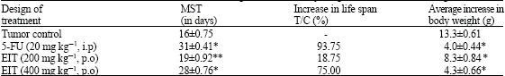 | |
| N = 6 animals in each group, Values are expressed as mean±SEM, *: p<0.001; **: p<0.01 when compared with control | |
Hematological parameters of tumor bearing mice on the day 14 were showed significant changes when compared to normal mice. The total WBC count, protein and PCV were found to increase with a reduction in the hemoglobin content of RBC. The differential count of WBC showed that the percentage of neutrophils increased while that of lymphocytes decreased (Table 2). At the same time interval, EIT (200 and 400 mg kg-1) treatment could change those altered parameters significantly (p<0.001; p<0.01) near to normal. Maximum alteration occurred in the EIT treatment at the dose of 400 mg kg-1.
There was significant reduction (p<0.001) in the tumor volume of mice treated with EIT on dose dependent manner. Tumor volume of control animals was increased by 14.2±0.38 mL, while it was 8.76±0.91 and 5.87±0.21 mL for the groups treated with EIT 200 and 400 mg kg-1, respectively (Table 3).
The reliable criteria for judging the value of any anticancer drugs are prolongation of life span, inhibition of gain in average body weight and decrease of WBC from blood (Clarkson and Burchenal, 1965; Obiling and Guerin, 1954). The results of the present study show the antitumour effect of EIT against EAC in mice. A significant (p<0.001) enhancement of MST and decrement of gain in average body weight was observed.
The analysis of the hematological parameters showed minimum toxic effect in mice treated with EIT. After 14 days of transplantation, EIT was able to reverse the changes in the hematological parameters consequent to tumor inoculation.
The common problems encountered in cancer chemotherapy are myelosuppression and anaemia (Marklund et al., 1982; Price and Greenfield, 1958). Anaemia occurring in tumour bearing mice is mainly due to reduction in RBC or haemoglobin production and this may occur either due to iron deficiency or due to haemolytic or other myelopathic conditions (Hogland, 1982). Treatment with EIT brought back the haemoglobin content, RBC and WBC counts to near normal. This indicates that EIT have a protective effect on the haemopoietic system. Further, analysis of haemotological parameters showed minimum toxic effect in mice treated with EIT. In EAC bearing mice, haematological parameters were reversed to normal by EIT administration (9 days).
In EAC bearing mice, there was a regular and rapid increase in ascitic fluid volume. Ascitic fluid is the direct nutritional source for tumour growth, it meets the nutritional requirement of tumour cells (Feng et al., 2001). EIT treatment decreased the volume of solid tumour, viable cancer cell count and increased the life span.
| Table 2: | Effect of EIT on hematological parameters of EAC-bearing mice |
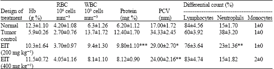 | |
| N = 5 animals in each group, Values are expressed as mean±SEM, *: p<0.001; **: p<0.01; ***: p<0.05 when compared with control | |
| Table 3: | Effect of EIT on solid tumor volume |
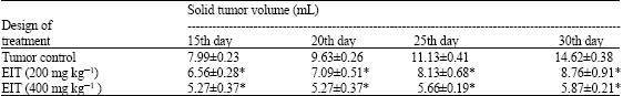 | |
| N = 6 animals in each group, Values are expressed as mean±SEM, *: p<0.001when compared with control | |
It may conclude that EIT decrease the nutritional fluid volume and thereby arrest the tumour growth and increase the life span. There was reduction in solid tumour volume of mice treated with EIT (p<0.001).
All these data point to possibly developing the ethanol extract of Indigofera trita as a novel and potential agent in the cancer chemotherapy. Preliminary phytochemical screening indicated the presence of alkaloids and flavonoids in EIT. Flavonoids have been shown to possess antimutagenic and antimalignant effects (Brown, 1980; Hirano et al., 1989). Moreover, flavonoids have a chemo preventive role in cancer through their effects on signal transduction in cell proliferation (Weber et al., 1996) and angiogenesis (Fotis et al., 1997). The antitumour properties of the extract may be due to these compounds. The present study points the potential anticancer activity of Indigofera trita in a dose dependent manner.
CONCLUSIONS
All these observations clearly indicate the significant antitumour effect of ethanol extract of Indigofera trita. Further studies to characterize the active principles and elucidate the mechanism of action of EIT are in progress.
REFERENCES
- Awadh Ali, N.A., W.D. Julich, C. Kusnick and U. Lindequist, 2001. Screening of Yemeni medicinal plants for antibacterial and cytotoxic activities. J. Ethnopharmacol., 74: 173-179.
CrossRefPubMedDirect Link - Brown, J.P., 1980. A review of the genetic effect of occurring flavonoids, anthraquinones and related compounds. Mut. Res., 75: 243-277.
PubMed - Christina, A.J., M. Alwin Jose, S.J. Heison Robert, R. Kothai, N. Chidambaranathan and P. Muthumani, 2003. Effect of Indigofera aspalathoides against Dalton`s ascitic lymphoma. Fitoterapia, 74: 280-283.
Direct Link - Feng, Q., T. Kumagai, Y. Torii, Y. Nakamura, T. Osawa and K. Uchida, 2001. Anticarcinogenic antioxidants as inhibitors against intracellular oxidative stress. Free Radical Res., 35: 779-788.
CrossRefPubMedDirect Link - Fotis, T., M.S. Pepper, E. Aktan, S. Breit and S. Rasku et al., 1997. Flavonoids, dietary derived inhibitors of cell proliferation and in vitro angiogenesis. Cancer Res., 57: 2916-2921.
Direct Link - Han, R., 1994. Highlights on the studies of anticancer drugs derived from plants in China. Stem Cells, 12: 53-63.
PubMed - Hoagland, H.C., 1982. Hematologic complications of cancer chemotherapy. Semin. Oncol., 9: 95-102.
PubMedDirect Link - Lowry, O.H., N.J. Rosebrough, A.L. Farr and R.J. Randall, 1951. Protein measurement with the folin phenol reagent. J. Biol. Chem., 193: 265-275.
CrossRefPubMedDirect Link - Marklund, S.L., N.G. Westman, E. Lundgren and G. Roos, 1982. Copper and zinc containing superoxide dismutase, manganese containing superoxide dismutase, catalase and glutathione peroxidase in normal and neoplastic human cell lines and normal human tissues. Cancer Res., 42: 1955-1961.
Direct Link - Newman, D.J., G.M. Cragg and K.M. Snader, 2003. Natural products as source of new drugs over the period 1981-2002. J. Nat. Prod., 66: 1022-1037.
PubMed - Price, V.E. and R.E. Greenfield, 1958. Anemia in cancer. Adv. Cancer Res., 5: 199-290.
PubMedDirect Link - Rajkapoor, B., B. Jayakar and N. Murugesh, 2004. Antitumor activity of Indigofera aspalathoides on Ehrlich ascites carcinoma in mice. Ind. J. Pharmacol., 36: 38-40.
Direct Link - Rajkapoor, B., N. Murugesh, D. Chodon and D. Sakthisekaran, 2005. Chemoprevention of N-nitrosodiethylamine induced phenobarbitol promoted liver tumors in rat by extract of Indigofera aspalathoides. Biol. Pharm. Bull., 28: 364-366.
PubMedDirect Link - Ramnath, V., G. Kuttan and R. Kuttan, 2002. Antitumour effect of abrin on transplanted tumours in mice. Indian J. Physiol. Pharmacol., 46: 69-77.
Direct Link - Weber, G., F. Shen, N. Prajda, Y.A. Yeh, H. Yang, Herenyiova and K.Y. Look , 1996. Increased signal transduction activity and down regulation in human cancer cells. Anticancer Res., 16: 3271-3281.
Direct Link