
Hayri Baba
Department of Biology, Faculty of Science and Arts, Celal Bayer University, Manisa, Turkey
International Journal of Botany
Year: 2008 | Volume: 4 | Issue: 3 | Page No.: 336-339


ABSTRACT
In this study, 3 new species and a new genus are recorded for the first time from Turkey. These records are genera of Prototrichia Rostaf., Prototrichia metallica (Berk.) Massee, Physarum confertum Macbr. and Stemonitis foliicola Ing., species. They have been isolated using the moist chamber technique.
PDF Abstract XML References Citation
How to cite this article
Hayri Baba, 2008. A New Myxomycetes Genus and Three Species Record for Turkey. International Journal of Botany, 4: 336-339.
DOI: 10.3923/ijb.2008.336.339
URL: https://scialert.net/abstract/?doi=ijb.2008.336.339
DOI: 10.3923/ijb.2008.336.339
URL: https://scialert.net/abstract/?doi=ijb.2008.336.339
INTRODUCTION
Myxomycetes, called plasmodial slime moulds or true slime moulds, are fungus-like organisms and characterized by an assimilative phase in the form of free living, multinucleate, acellular, mobile mass of protoplasm, in a propagative phase, consisting of a mass of spores borne in a simple or complex, membranous, noncellular spore case. The members of the group are widely distributed, occurring wherever conditions on the earth`s surface permit the growth of vegetation, but are particularly abundant in the forested areas, where they appear in great profusion on dead and decaying wood, or woody litter and on dead leaves (Martin and Alexopoulos, 1969).
The number of myxomycete taxa known world wide is about 1000 (Ergul and Oran, 2005), but in Turkey, only about 216 taxa have been reported (Yağiz and Afyon, 2007). For these reasons, there is a great need for an increase of the frequency of taxonomical and mycofloristical studies. With the increased number of similar studies, it will be possible to fill in the gap and also it may cause a rise in the new myxomycetes taxa for Turkey.
Related studies (Lado, 1994; Ergül and Dülger, 2000, 2002a, b; Ocak and Hasenekoğlu, 2003, 2005; Sesli and Denchev, 2005; Demirel et al., 2006; Yağiz and Afyon, 2007) were reviewed and it was found that new taxa have been found for Turkey. With this study a new myxomycetes genus and three species were added to the Turkish myxomycetes flora as new records.
MATERIALS AND METHODS
The samples were collected from the Manisa in 2006. The myxomycetes fructification has been obtained by using of the moist chamber technique in the laboratory. Several kinds of plant remains and bark from living trees were kept in an incubator at the temperature of 25±0.1°C illuminated artificially in a 12:12 h light:dark cycle. The cultures were moistened with sterile distilled water adjusted with KOH to pH: 7.0. After two days the pH of the moisture in the dishes was measured with pH sticks. The moist chamber was then examined every second or third days under a dissecting microscope. When developing myxomycetes were found, the moist chamber was allowed to dry slowly and the myxomycetes were the dried for a week. Entire chamber was then rewetted for another four-week period and examined as before (Gilbert and Martin, 1933; Harkonen and Uotila, 1983).
The specimens were preserved also as permanent slides in Hoyer`s medium. Both microscopic and macroscopic observations have been realized for taxonomical approaches. In the meantime, some photographs from characteristics qualitative objects are taken. All data have been evaluated comparatively for taxonomical purposes (Martin and Alexopoulos, 1969).
The myxomycetes specimens were identified with the aid of the literature listed in the references (Martin and Alexopoulos, 1969; Neubert et al., 1993, 1995, 2000; Stephenson and Stempen, 2000). These specimens are stored at the Herbarium of Celal Bayar University, Manisa, Turkey.
RESULTS AND DISCUSSION
Totally, a genus and three species found to be new record for Turkey. These records are genera of Prototrichia Rostaf. and Prototrichia metallica (Berk.) Massee, Physarum confertum Macbr. and Stemonitis foliicola Ing., species. These taxa are explained in below.
A. Taxonomic position
| Regnum | : | Myceteae |
| Division | : | Gymnomycota |
| Class | : | Myxomycetes |
| Subclass | : | Myxogastromycetidae |
| Order | : | Trichiales |
| Family | : | Trichiaceae |
| Genus | : | Prototrichia Rostaf. Mon. App.38;1876. |
Description: Prototrichia metallica (Berk.) Massee, Jour. Roy. Micr. Soc. 350; 1889.
Syn: Trichia metallica Berk. In: Hooker f., Fl. Tasm. 2: 268; 1859.
Sessile or rarely short-stalked sporangia, scattered to gregarious, globose to subglobose, orange-brown to dull brown or sometimes rosaceous, 0.5 to 2.2 mm in diameter; peridium thin, transparent, often somewhat iridescent; capillitium consisting of numerous yellow-brown, more or less spirally banded threads that originate at the base of the sporangium and ultimately become somewhat subdivided above; capillitial threads fairly stout, twisted around each other in bundles, usually marked at least in part with faint to distinct spiral bands (nearly smooth elsewhere); spores orange brown to dull brown in mass, yellow by transmitted light, spiny, 12 to 15 μm in diameter (Fig. 1).
Locality: Demirci - Bardakçi village, Cevizdere area, on decaying wood of Pinus nigra, the altitude is 1050 m. 18.06.2006, H. Baba.
B. Taxonomic position
| Regnum | : | Myceteae |
| Division | : | Gymnomycota |
| Class | : | Myxomycetes |
| Subclass | : | Myxogastromycetidae |
| Order | : | Physarales |
| Family | : | Physaraceae |
| Genus | : | Physarum |
Description: Physarum confertum Macbr. T. Macbr. N. Am. Slime Moulds ed.2:64; 1922.
Always sessile sporangia, 0.2-0.8 mm length, sporangia gregarious, crowded together, globose or some sporangia depressed globose, slightly umbilicate at base, white to black or sometimes grey brown, 0.2 mm diameter; peridium covered with white-gray lime crystals; columella absent; capillitium delicate, branched and anastomosed threads, hyaline and colourless; spores brown in mass, pale violaceous in transmitted light, globose, fairly warted, 12-14 μm in diameter (Fig. 2).
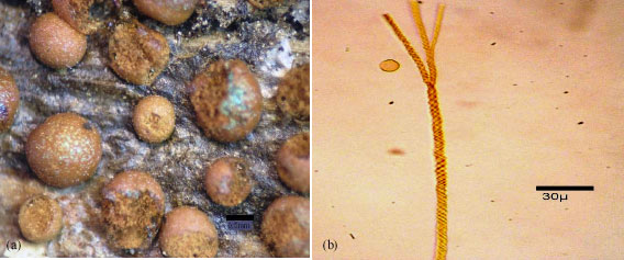 | |
| Fig. 1: | (a) Stereomicroscopic image of the sporangia of Prototrichia metallica (Berk.) Massee, (b) Spores and capillitium |
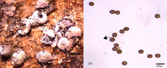 | |
| Fig. 2: | (a) Stereomicroscopic image of the sporangia Physarum confertum Macbr., (b) Spores and capillitium |
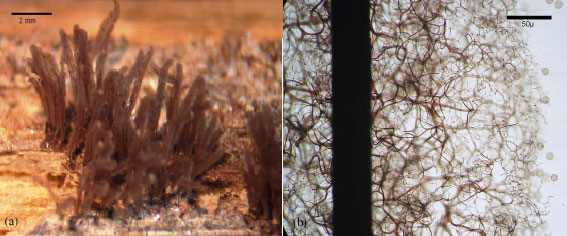 | |
| Fig. 3: | (a) Stereomicroscopic image of the sporangia of Stemonitis foliicola Ing., (b) Spores, capillitium and columella |
Locality: Manisa - Laleli, on decaying wood of Salix sp. the altitude is 85 m. 10.12.2006, H. Baba.
C. Taxonomic position
| Regnum | : | Myceteae |
| Division | : | Gymnomycota |
| Class | : | Myxomycetes |
| Subclass | : | Stemonitomycetidae |
| Order | : | Stemonitales |
| Family | : | Stemonitaceae |
| Genus | : | Stemonitis |
Description: Stemonitis foliicola Ing. Trans. Brit. Mycol. Soc.50: 555;1967.
Sporophor sporangiate, stipitate, densely clustered, cylindrical narrowed at upper part, 0.6-0.8 mm. in diameter, 3-4 mm. in length, dark brown; hypothallus dark brown, reddish brown in transmitted light; stipe bright olivaceous black colours, 1-1.5 mm in length; peridium evanescent but persists as scattered small flakes, remnants at upper parts of sporangia; columella black in transmitted light; capillitium dark brown, arising from the entire columella, primary branches stout, bearing numerous expansions, branched and anastomosed, arising the delicate small meshed peridial net, meshes mostly 10-15 μm in diameter, free ends numerous; spore mass dark brown, red in transmitted light, verrucose-reticulate, 7-9 μm in diameter, there are 6-9 border in a spore (Fig. 3).
Locality: Spil Mountain, Sultan plateau, on debris of Pinus nigra, the altitude is 850 m. 04.09.2005, H. Baba; Sarigöl - Karacaali village, on piece of branch of Quercus sp. the altitude is 720 m. 25.07.2006, H. Baba.
Since, Gilbert and Martin (1933) accidentally discovered that the moist chamber technique was an excellent method for obtaining fructifications of the standard means for studies and inventories of myxomycetes. It is applicable to almost any kind of substrate but has often been used for bark samples. Moist chamber cultures have yielded sporangia of species too small to be detected in the field and several new species have been described (Eliasson, 1991).
Prototrichia metallica (Berk.) Massee is one of the more distinctive snowbank myxomycetes characteristically found in alpine regions of the temperate zone, cosmopolitan and common, especially on decaying wood and bark of coniferous trees (Gilbert and Martin, 1933). We found this species on wood of Pinus nigra.
Physarum confertum Macbr. varies from other species of Physarum Pers. by it has always sessile sporangia, sporangia gregarious, crowded together, sporangia depressed globose, slightly umbilicate at base (Neubert et al., 2000). Our samples show the same characteristics. Our sporangia limy iridescent at base.
Stemonitis foliicola Ing. can be distinguished from species of Stemonitis by its peridium evanescent but persists as scattered small flakes, remnants at upper parts of sporangia, sporangia narrowed at upper part, delicate small meshed capillitial net, meshes mostly 10-15 μm in diameter, free ends numerous; spore verrucose-reticulate, 7-9 μm in diameter, there are 6-9 border in a spore.
In conclusion, the number of known Myxomycetes species in the world is 1000 only about 100 taxa have been reported with the moist chamber technique and naturally in Turkey (Ergul and Oran, 2005; Dulger et al., 2005).
REFERENCES
- Demirel, G., G. Kaşık and C. Öztürk, 2006. Myxomycetes of Kestel forest (Kadınhanı, Konya). Turk. J. Bot., 30: 441-447.
Direct Link - Dulger, B., E. Karabacak, T.B. Suerdem and N. Hacioglu, 2005. A new myxomycete record for the fungi flora of Turkey. Int. J. Bot., 1: 62-63.
Direct Link - Eliasson, U.M., 1991. The myxomycete biota of the Hawaiian Islands. Mycol. Res., 95: 257-267.
CrossRefDirect Link - Ergul, C.C. and B. Dulger, 2002. A new record for the myxomycetes flora of Turkey: Comatricha pulchella (C.Bab.) Rost. var. pulchella. Turk. J. Bot., 26: 113-115.
Direct Link - Ergul, C.C. and B. Dulger, 2002. New record for the myxomycetes flora of Turkey. Turk. J. Bot., 26: 277-280.
Direct Link - Ergul, C.C. and R.B. Oran, 2005. Three new records for the Turkish Myxobiota. Turk. J. Bot., 29: 241-242.
Direct Link - Harkonen, M. and P. Uotila, 1983. Turkish myxomycetes developed in moist chamber cultures. Karstenia, 23: 1-9.
Direct Link - Lado, C.A., 1994. Cheklist of myxomycetes of the Mediterranean countries. Mycotaxon, 52: 117-185.
Direct Link - Ocak, İ. and İ. Hasenekoglu, 2003. Myxomycetes from Erzurum, Bayburt and Gümüşhane provinces (Turkey). Turk. J. Bot., 27: 223-226.
Direct Link - Ocak, İ. and İ. Hasenekoğlu, 2005. Myxomycetes from Trabzon and Giresun provinces (Turkey). Turk. J. Bot., 29: 11-21.
Direct Link - Sesli, E. and C.M. Denchev, 2005. Checklists of the myxomycetes and macromycetes in Turkey. Mycol. Balcanica, 2: 119-160.
Direct Link