
C. Zongo
Centre de Recherche en Sciences Biologiques, Alimentaires et Nutritionnelles, UFR/SVT, Universite de Ouagadougou, 03 BP 7131, Ouagadougou 03, Burkina Faso
Etienne-Francois O. Akomo
Centre de Recherche en Sciences Biologiques, Alimentaires et Nutritionnelles, UFR/SVT, Universite de Ouagadougou, 03 BP 7131, Ouagadougou 03, Burkina Faso
A. Savadogo
Centre de Recherche en Sciences Biologiques, Alimentaires et Nutritionnelles, UFR/SVT, Universite de Ouagadougou, 03 BP 7131, Ouagadougou 03, Burkina Faso
Louis Clement Obame
Centre de Recherche en Sciences Biologiques, Alimentaires et Nutritionnelles, UFR/SVT, Universite de Ouagadougou, 03 BP 7131, Ouagadougou 03, Burkina Faso
Jean Koudou
Centre de Recherche en Pharmacopee et Medecine Traditionnelle, Universite de Bangui, BP 1450, Bangui Republique Centrafricaine
Alfred S. Traore
Centre de Recherche en Sciences Biologiques, Alimentaires et Nutritionnelles, UFR/SVT, Universite de Ouagadougou, 03 BP 7131, Ouagadougou 03, Burkina Faso
Asian Journal of Plant Sciences
Year: 2009 | Volume: 8 | Issue: 2 | Page No.: 172-177


ABSTRACT
The antimicrobial activity of the total alkaloids from the leaves of Mitragyna inermis (Willd.) O. Kuntze (Rubiaceae) From Burkina Faso was evaluated using disc diffusion essay and broth microdilution assay. The extract was used against ten (10) reference bacterial strains and three (3) clinical isolates including Gram(+) and Gram(-) strains. The alkaloids showed moderate activity against mcroorganisms tested. The highest Diameter of Inhibition Zone (DIZ) was obtained with S. aureus ATCC9244 (23 ± 1 mm). The lowest Minimum Inhibitory Concentration (MIC) obtained was 0.625 mg mL-1 recorded with three (3) Gram(+) bacteria (S. aureus ATCC 25293, S. aureus (clinical isolate) and S. carmorum LMG 13567) and one Gram(-) strain (P. mirabilis CIP104588. Results showed that Gram(+) bacteria are more sensitive to alkaloids from M. inermis than Gram(-) bacteria. This study confirmed the use of the plant in traditional medicine against some infectious diseases.
PDF Abstract XML References Citation
How to cite this article
C. Zongo, Etienne-Francois O. Akomo, A. Savadogo, Louis Clement Obame, Jean Koudou and Alfred S. Traore, 2009. In vitro Antibacterial Properties of Total Alkaloids Extract from Mitragyna Inermis (Willd.) O. Kuntze, a West African Traditional Medicinal Plant. Asian Journal of Plant Sciences, 8: 172-177.
DOI: 10.3923/ajps.2009.172.177
URL: https://scialert.net/abstract/?doi=ajps.2009.172.177
DOI: 10.3923/ajps.2009.172.177
URL: https://scialert.net/abstract/?doi=ajps.2009.172.177
INTRODUCTION
Indigenous medicinal plants are often the only means for the treatment of several infections in Africa (Fennell et al., 2004; Taylor et al., 2001). According to WHO (2003) 80% of the population use traditional medicine for their primary health care.
Mitragyna inermis (Willd.) O. Kuntze (Rubiaceae) is a medicinal plant widely known and used in folk medicine in West Africa. This plant is a shrub growing on low alluvial plains and swampy savannah of many countries of West Africa (Pillay, 1964; Shellard and Wade, 1969; Kerharo Adam, 1974).
In traditional medicine, M. inermis is used to treat several diseases such as fever, headache, diarrhoea, dysentery, cholera, malaria and other diseases. (Nacoulma/Ouèdraogo, 1996; Ouedraogo et al., 2007). According to the traditional knowledge, many scientific studies have been carried out to confirm the activities the plant is assumed to exert in vivo.
The in vitro antiplasmodial activity of the plant has been demonstrated by Traore-Keita et al. (2000), Mustofa et al. (2000), Kohler et al. (2002), Azas et al. (2002) and Fiot et al. (2005). The cardiovascular properties of the aqueous extract have been also demonstrated (Ouedraogo et al., 2004). Toxicity studies including cytotoxicity, genotoxicity, acute and chronic toxicity have been carried out (Azas et al., 2002; Toure et al., 1996; Traore et al., 2000; Monjanel-Mouterde et al., 2006). Several compounds including alkaloids and non-alkaloids have been purified from M. inermis (Shellard and Sarpong, 1969, 1970; Shellard et al., 1971; Cheng et al., 2002; Fiot et al., 2005). The chloroformic extract from the plant showed antibacterial activity against Escherichia coli, Staphylococcus aureus and Streptococcus sp. (Umeh et al., 2005). Some studies showed that the pharmacological and biological properties were mostly due to alkaloids (Traore-Keita et al., 2000). More recently, antimicrobial activity of different extracts from M. inermis against bacteria and fungi has been demonstrated and chemical analysis revealed the presence of alkaloids in the active extracts (Asase et al., 2008).
However, there is a few scientific data on the antibacterial properties of the plant. In the present study, in vitro antibacterial activity of the total alkaloids of M. inermis was evaluated against clinical and reference Gram-positive and Gram-negative bacteria.
MATERIALS AND METHODS
Plant material: The leaves of Mitragyna inermis were collected in December 2007, at 45 km from Ouagadougou in Burkina Faso. The samples were carefully dried in the laboratory under continuous ventilation, away from sun light and dust. The leaves were then crushed to fine powder with a mechanical crusher and the powder was kept in plastic bags and stored away from light and moisture until required.
The plant was taxonomically authenticated at the Laboratory of Plant Biology and Ecology of the University of Ouagadougou where a voucher specimen was deposited.
Total alkaloids extraction: The dried powder of the leaves was moistened with ammonia (28%) and extracted with chloroform at room temperature for a total period of 24 h. The chloroform extract was then filtered and a first liquid-liquid partition of the alkaloids was made with hydrochloric acid (HCl) (5%). The aqueous layer from the first partition was made alkaline again (pH 9-10) with ammonia and a second partition with chloroform was made. Finally, chloroform was totally evaporated from the organic phase to give a total alkaloids powder.
Microbial strains: The total alkaloids extract of leaves of M. inermis was tested against a panel of microorganisms, including reference strains (Bacillus cereus LMG 13569, Enterococcus faecalis CIP 103907, Escherichia coli CIP 105182, Listeria innocua LMG 1135668, Salmonella enterica CIP 105150, Shigella dysenteria CIP 5451, Staphylococcus aureus ATCC 9244, Proteus mirabilis CIP 104588 Staphylococcus aureus ATCC 25293 and Staphylococcus camorum LMG 13567) and Clinical strains (Pseudomonas aeruginosa, Staphylococcus aureus and Streptococcus pyogenes). These clinical strains were isolated at the Laboratoire de Biologie Médicale Saint Camille in Ouagadougou. The identification of these clinical strains was based on their biochemical profiles as recommended by the manual Bactériologie Médicale (Le-Minor and Veron, 1984).
Antibiotics and media: Commercially available antibiotics discs, ampicillin 33 μg and Tetracyclin 30 μg were purchased from Beckton Dickinson and used as references for the test. All media used were from Fluka BioChemica. Chloroform was analytical grade.
Antibacterial assays
Disc diffusion method: The in vitro antibacterial activity of total alkaloids extract of M. inermis was studied by the paper disc diffusion method (Bauer et al., 1966; Pelczar et al., 1993; Ayandele and Adebiyi, 2007) using Mueller-Hinton agar plates. Briefly, to activate the microorganisms, they were grown on nutrient broth at 37 °C for 18 h. The other night cultures were suspended in saline solution (0.9% NaCl) and adjusted to a turbidity of 0.5 Mac Farland standards corresponding to 108 cfu mL-1. Each bacterial suspension was used to inoculate 90 mm diameter Petri plates with a sterile non toxic cotton swab. Six millimeter paper discs (Whatman No. 3) soaked with 10 μL of the total alkaloid extract dilution (100 mg mL-1 in dimethylsulphoxyde (DMSO) were placed on the agar. The quantity of extract was then 1000 μg per disc. Paper discs soaked in DMSO without extract were used as negative control and DMSO didn’t show inhibition effects to mcroorganisms growth. The plates were incubated aerobically at 37 °C for 18 to 24 h. All tests were performed in duplicate and the antibacterial activity was expressed as the mean of Diameters of Inhibition Zone (DIZ) produced.
Broth microdilution assay: A microdilution broth susceptibility assay was used, as recommended by the National Committee for Clinical Laboratory Standards (2006) for the determination of the Minimum Inhibitory Concentration (MIC) and the minimum bactericidal concentration (MBC). Briefly, the total alkaloids extract was properly prepared, sterilized by filtration through 0.22 μm sterilizing Millipore express filter and transferred in sterile 96 well-plates previously filed with sterile nutrient broth to obtain a twofold serial dilutions ranging from 19,5312 to 2500 μg mL-1. Then plates were inoculated with microbial suspensions diluted from the same 0.5 Mac Farland standards to have 5x10⁵ cfu mL-1 in each well. A number of wells were reserved in each plate for sterility control (no inoculum added), inoculum viability (no extract added) and the DMSO inhibitory effect. The final volumes in wells were 200 μL. After 24 h aerobically incubation at 37 °C, bacterial growth was indicated by the presence of turbidity and a pellet on the well bottom. MIC was defined as the lowest extract concentration demonstrating no visible growth in the broth and the MBC was defined as the lowest concentration of the extract at which 99.99% or more of the initial inoculum was killed. To determine MBC values, 100 μL of bacterial suspension from subculture demonstrating no visible growth were removed to spread onto Plate Count Agar (PCA) medium plates. Plates were incubated at 37 °C for a total period of 48 h.
Time-kill assay: In order to evaluate the efficiency of the alkaloids as a function of the time, two bacteria (E. coli CIP 105182 and S. enterica CIP 105150) were arbitrary chosen to perform a time-kill assay according to NCCLS guidelines (National Committe for Clinical Laboratory, 1999). For this test, other night bacteria cultures were adjusted to the same 0.5 Mac Farland standard and then diluted in 50 mL of sterile nutrient broth to have approximately 105 cfu mL-1. The alkaloids were added at the concentration of 3 mg mL-1 for both bacteria. This concentration chosen arbitrary was slightly greater than their MBC values. The cultures were incubated aerobically at 37 °C in an incubator shaker (INNOVATM 4000). Each hour, an aliquot of 100 μL was removed from each culture and diluted with 10 mL sterile isotonic water. Successive dilutions were made from this initial dilution and the obtained suspension was used to inoculate 90 mm diameter Petri plates previously filed with PCA (Plate Count Agar). After 48 h incubation at 37 °C, The number of viable bacteria was evaluated by Colonies Forming Units (CFU) counting. The experiment was carried out twice.
RESULTS AND DISCUSSION
The aim of this research was to study the antibacterial activity of total alkaloids of M. inermis. Present results exhibited moderate to significant antibacterial activity against bacterial strains used for this test. The diameters of inhibition zone (DIZ) indicated the susceptibility of all tested bacteria to alkaloids excepted to P. aeruginosa. An antibacterial activity is recorded when a DIZ more than 9 mm is observed around the paper disc (Kitzberger et al., 2006). The largest DIZ (23 ± 1) mm were obtained with S. aureus ATCC92449. It has frequently been reported that Gram-positive bacteria are more sensitive to plant extract and their components than Gram-negative bacteria (Kelmanson et al., 2000; Masika and Afolayane, 2002 ; Sahin et al., 2002; Karaman et al., 2003; Karou et al., 2006; Masoodi et al., 2008). The results of this study confirmed these observations. Diameter of inhibition values obtained with gram-positive bacteria are larger than those obtained with gram-negative bacteria (Table 1). Earlier study with alkaloids of Sida acuta (Karou et al., 2006) gave DIZ values greater than those obtained in this study. However, it is difficult to make a comparison because alkaloids from M. inermis are different from those from S. acuta. It is also indicated that the DIZ value is determined by the initial population density of the microorganisms, their growth rate and the rate of diffusion of the antimicrobial agent (Hugo and Russell, 1998).
The lowest MIC (0.625 mg mL-1) was recorded with P. mirabilis CIP104588, S. aureus (clinical isolate), S. carmorum LMG13567 and S. aureus ATCC 25293. The antibacterial activity was considered bactericide when the ratio MBC/MIC is 1 or 2 and bacteriostatic when this ratio is 3 or more. In this case, alkaloids of M. inermis can be considerate bactericide to tested bacteria (Table 2). The mode of action of the alkaloids of M. inermis which include indole and oxindole alkaloids (Shellard and Sharpong, 1969) is not known in detail. They should have a particular mechanism which can justified their actions against Gram negative and Gram positive bacteria because the outer membrane of Gram-negative bacteria is known to present a barrier to the penetration of numerous antibiotic molecules.
| Table 1: | Diameters of inhibition zone by disc diffusion assay |
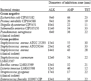 | |
| ALK: Alkaloids of M. inermis; AMP: Ampicillin; TET: Tetracyclin | |
| Table 2: | MIC and MBC values of total alkaloids of M. inermis in the microdilution assay |
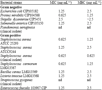 | |
| nd: Not determine | |
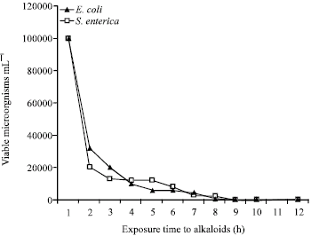 | |
| Fig. 1: | Evolution of bacteria exposed to alkaloids (3 mg mL-1) in the time-kill assay |
It have been reported that alkaloids of the quinolone family act by targeting bacterial DNA synthesis essential enzymes such as DNA gyrase and DNA topoisomerase IV, that play important roles in DNA replication (Drica and Zhao, 1997; Khodursky and Cozzarelli, 1998) or competitively inhibit electron transport in the respiratory chain (Dekker et al., 1998; Kunze et al., 1987). Other studies have shown that bisindole monoterpenoid alkaloids act as DNA intercalating agents or like topoisomerase inhibitors (Angenot et al., 1991; Bonjean et al., 1998).
In the time-kill assay performed to follow the alkaloids on microorganisms as a function of the time, both test bacteria (E. coli CIP 105182 and S. enterica CIP 105150) showed the same clearance time (Fig. 1). After 9 h exposition, any CFU was observed in the plates traducing that there was no viable microorganism in the initial inoculums.
In conclusion, the study has shown that alkaloids from M. inermis also have in vitro antimicrobial activities, which could support the use of the plant by traditional healers to treat various infective diseases Further studies could led to the most active alkaloids and could led to a new antimicrobial agent.
ACKNOWLEDGMENTS
This study was partially supported by CORUS 034. We gratefully think Dr. Imaël H.N. Bassole for his helpful comments on the manuscript and technical help and Mr. Dieudonné Kinda for his help during plant material collection. We also think Laboratoire de Biologie Médicale Saint Camille for providing clinical bacterial strains.
REFERENCES
- Angenot, L., J. Quentin-Leclercq, D.J. Phillipson, D.C. Warhurst, M.J. O'Neill, D.H. Bray and C.W. Wright, 1991. Antiamoebic and antiplasmodial activities of alkaloids isolated from Strychnos usambarensis. Planta Med., 57: 337-340.
PubMed - Asase, A., T. Kokubun, R.J. Grayer, G. Kite, M.S.J. Simmonds, A.A.O. Yeboah and G.T. Odamtten, 2008. Chemical constituents and antimicrobial activity of medicinal plants from Ghana: Cassia sieberiana, Haematostaphis barteri, Mitragyna inermis and Pseudocedrela kotschyi. Phytother. Res., 22: 1013-1016.
CrossRefPubMedDirect Link - Bauer, A.W., W.M.M. Kirby, J.C. Sherris and M. Turck, 1966. Antibiotic susceptibility testing by a standardized single disk method. Am. J. Clin. Pathol., 45: 493-496.
CrossRefPubMedDirect Link - Bonjean, K., M.C. De Pauw-Gillet, M.P. Defresne, P. Colson and C. Houssier et al., 1998. The DNA intercalating alkaloid cryptolepine interferes with topoisomerase II and inhibits primarily DNA synthesis in B16 melanoma cells. Biochemistry, 37: 1536-1546.
CrossRef - Drlica, K. and X. Zhao, 1997. DNA gyrase, topoisomerase IV and the 4-quinolones. Microbiol. Mol. Biol. Rev., 61: 377-392.
PubMedDirect Link - Dekker, K.A., T. Inagaki, T.D. Gootz, L.H. Huang and Y. Kojima et al., 1998. New quinolone compounds from Pseudonocardia sp. with selective and potent anti-Helicobacter pylori activity: Taxonomy of producing strains, fermentation, isolation, structural eradication and biological activities. J. Antibiot., 51: 145-152.
PubMed - Fennell, C.W., K.L. Lindsey, L.J. McGaw, S.G. Sparg and G.I. Stafford et al., 2004. Assessing African medicinal plants for efficacy and safety: Pharmacological screening and toxicology. J. Ethnopharmacol., 94: 205-217.
CrossRefPubMedDirect Link - Karaman, I., F. Sahin, M. Gulluce, H. Ogutcu, M. Sengul and A. Adiguzel, 2003. Antimicrobial activity of aqueous and methanol extracts of Juniperus oxycedrus L. J. Ethnopharmacol., 85: 231-235.
CrossRefPubMedDirect Link - Karou, D., A. Savadogo, A. Canini, S. Yameogo and C. Montesano et al., 2005. Antibacterial activity of alkaloids from Sida acuta. Afr. J. Biotechnol., 4: 1452-1457.
Direct Link - Kelmanson, J.E., A.K. Jager and J. van Staden, 2000. Zulu medicinal plants with antibacterial activity. J. Ethnopharmacol., 69: 241-246.
CrossRefPubMedDirect Link - Khodursky, A.B. and N.R. Cozzarelli, 1998. The mechanism of inhibition of topoisomerase IV by quinolone antibacterials. J. Biol. Chem., 273: 27668-27677.
PubMed - Kitzberger, C.S.G., A. Smania, R.C. Pedrosa and S.R.S. Ferreira, 2006. Antioxidant and antimicrobial activities of shiitake (Lentinula edoles) extracts obtained by organic solvents and supercritical fluids. J. Food Eng., 80: 631-638.
CrossRef - Köhler, I., K. Jennet-Siems, K. Carola, K. Siems, D. Abbiw, U. Bienzle and E. Eich, 2002. Herbal remedies traditionaly used against malaria in Ghana: Bioassay-guided fractionation of Microglossa pyrifolia (Asteraceae). Z. Naturforsch., 57c: 1022-1027.
PubMed - Kunze, B., G. Hoflea and H. Reichenbach, 1987. The aurachins, new quinolone antibiotics from myxobacteria: Production, physico-chemical and biological properties. J. Antibiot., 40: 258-265.
PubMed - Masoodi, M.H., B. Ahmed, I.M. Zargar, S.A. Khan, S. Khan and P. Singh, 2008. Antibacterial activity of whole plant extract of Marrubium vulgare. Afr. J. Biotechnol., 7: 86-87.
Direct Link - Masika, P.J. and A.J. Afolayane, 2002. Antimicrobial activity of some plants used for the treatment of livestock diseases in Eastern Cape, South Africa. J. Ethnopharmacol., 83: 129-134.
CrossRef - Monjanel-Mouterde, S., F. Traora, M. Gasquet, F. Dodero and F. Delmas et al., 2006. Lack of toxicity of hydroethanolic extract from Mitragyna inermis (Willd.) O. Kuntze by gavage in the rat. J. Ethnopharmacol., 103: 319-326.
PubMed - Ouedraogo, S., H.R. Ranaivo, M. Ndiaye, Z.I. Kabore, I.P. Guissou, B. Bucher and R. Andriantsitohaina, 2004. Cardiovascular properties of aqueous extract from Mitragyna inermis (Wild). J. Ethnopharmacol., 93: 345-350.
PubMed - Ouedraogo, Y., I.P. Guissou and O.G. Nacoulma, 2007. Biological study of aqueous root extract from Mitragyna inermis (WILLD oktze) Rubiaceae. Int. J. Pharmacol., 3: 80-85.
Direct Link - Pillay, M.J., 1964. Anatomy of leaves and young stem of Mitragyna inermis (O. Kuntze). J. Pharm. Pharmacol., 16: 820-827.
PubMed - Sahin, F., I. Karaman, M. Gulluce, H. Ogutcu and M. Sengul et al., 2003. Evaluation of antimicrobial activities Satureja hortensis L. J. Ethnopharmacol., 87: 61-65.
CrossRef - Shellard, E.J. and K. Sharpong, 1969. The alkaloids of the leaves of Mitragyna inermis (Willd.) O. Kuntze. J. Pharm. Pharmacol., 21: 113S+-113S+.
PubMed - Shellard, E.J. and A. Wade, 1969. The morphology and anatomy of the flowers of Mitragyna inermis (Willd.) O. Kuntze. J. Pharm. Pharmacol., 21: 102S-112S.
PubMed - Shellard, E.J. and K. Sarpong, 1970. The alkaloidal pattern in the leaves, stem-bark and root-bark of Mitragyna species from Ghana. J. Pharm. Pharmacol., 22: 34S+-34S+.
PubMed - Shellard, E.J., J.D. Phillipson and K. Sarpong, 1971. N-Oxides of the oxindole alkaloids rhynchophylline and isorhynchophylline. Xenobiotica, 1: 455-456.
CrossRef - Taylor, J.L.S., T. Rabe, L.J. McGaw, A.K. Jager and J. van Staden, 2001. Towards the scientific validation of traditional medicinal plants. Plant Growth Regul., 34: 23-37.
CrossRefDirect Link - Toure H., G. Balansard, A.M. Pauli and A.M. Scotto, 1996. Pharmacological investigation of alkaloids from leaves of Mitragyna inermis (Rubiaceae). J. Ethnopharmacol., 54: 59-62.
PubMed - Traore, F., M. Gasquet, M. Laget, H. Guiraud and C. Di-Giorgio et al., 2000. Toxicity and genotoxicity of antimalarial alkaloid rich extracts derived from Mytragyna inermis O. Kuntze and Nauclea latifolia. Phytother. Res., 14: 608-611.
PubMed - Traore-Keita, F., M. Gasquet, C. Di Giorgio, E. Ollivier and F. Delmas et al., 2000. Antimalarial activity of four plants used in traditional medicine in Mali. Phytother. Res., 14: 45-47.
CrossRefPubMedDirect Link - Umeh, E.U., H.O.A. Oluma and J.O. Igoli, 2005. Antibacterial screening of four local plants using an indicator-based microdilution technique. Afr. J. Trad. Compl. Alter. Med., 2: 238-243.
Direct Link
ahmad shafiei darabi Reply
Dear
I am ahmad shafiei
I graduated from shahid beheshti university, Iran
I work on galbanum and have a paper about it.
The title of paper is "The comparison of galbanum (Ferula gummosa Boiss) from five regions of Iran"
Pleas send your address or email
Thanks
zongo cheikna
Thank you for you interest for my work. You can make contact with me for scientic exchanges and over.
Best regards!
saeed aria Reply
hello i am horticulture engineerig student in tehran!
i"d look like
your aricle was good