
M.A. Eroje
Department of Anatomy, Niger Delta University, Wilberforce Island, Amassoma, Bayelsa State, Nigeria
F.M. Onyije
Department of Anatomy, Niger Delta University, Wilberforce Island, Amassoma, Bayelsa State, Nigeria
Asian Journal of Biological Sciences
Year: 2011 | Volume: 4 | Issue: 2 | Page No.: 196-200


ABSTRACT
The lung which is a vital organ of respiration normally has two lobes on the left and three on the right with the left lung having an oblique fissure and the right having both an oblique and a horizontal fissure. Although, many clinicians have observed variations in lobar pattern with the aid of CT scans, only few studies on gross anatomical specimens have been reported. In this report, we present variation in two right lungs as seen in the Gross Anatomy Laboratory of the College of Health Sciences, Niger Delta University (NDU), Bayelsa State, Nigeria. The first has a horizontal fissure and an incomplete oblique fissure while the second has a complete oblique fissure with a complete absence of the horizontal fissure. The variation in fissures and lobes presented in this report are clinically important in appreciating lobar anatomy and for identifying broncho-pulmonary segments. Knowledge of anatomical variations is essential for interpreting radiological images, performing lobectomies and also for general knowledge to medical personnel.
PDF Abstract XML References Citation
How to cite this article
M.A. Eroje and F.M. Onyije, 2011. Lobar Variation of Right Lung as Seen in a Nigerian Cadaver: A Case Report. Asian Journal of Biological Sciences, 4: 196-200.
URL: https://scialert.net/abstract/?doi=ajbs.2011.196.200
URL: https://scialert.net/abstract/?doi=ajbs.2011.196.200
INTRODUCTION
Lobar anatomy and bronchioplumonary segments can be appreciated better with knowledge of variations that abound in fissures of the lungs. Anatomically, the right lung has two fissures, an oblique and a horizontal dividing it into three lobes namely the superior, middle and lower (Standring, 2005). The oblique fissure cuts the vertebral border of both the lungs at the level of 4th or 5th thoracic spine. Traced downwards on the medial surface it ends above the hilum; traced downwards on the costal surface, it will be found to continue across the diaphragmatic surface and turn upward on to the medial surface to end just below the lower end of the hilum (Rosse and Gaddum-Rosse, 1997). The horizontal fissure, which is seen only in the right lung passes from the anterior margin into the oblique fissure to separate a wedge-shaped middle lobe from the upper lobe (Last, 2000). It passes at the level of midaxillary line to the anterior border of the lung at the level of sternal end of 4th costal cartilage (Standring, 2005).
The right lung is larger and heavier than the left, but it is shorter and wider because the right dome of the diaphragm is higher and the heart and pericardium bulge more to the left (Moore and Dally, 2006). The fissures may be complete, when the lobes remain held together only at the hilum by the bronchi and pulmonary vessels, or they may be incomplete when there are areas of parenchymal fusion between the lobes, or, they may be absent altogether (Meenakshi et al., 2004).
The knowledge of anatomical variations of the lobes of the lung is important for identifying broncho-pulmonary segments. Many a times, radiologists may misinterpret an x ray or CT scan. Anatomical knowledge of such variations is helpful for lobectomies and surgical resections involving individual segments (Modgil et al., 2006).
CASE REPORT
During routine dissection of the thoracic region in the dissecting laboratory of the Department of Anatomy, Niger Delta University in 2009, we encountered two anomalous right lungs which displayed variation in the pattern of fissures and lobes. There was no history of pulmonary disease in the individual. The lobes and the fissures were observed and the lungs were photographed (Fig. 1-5).
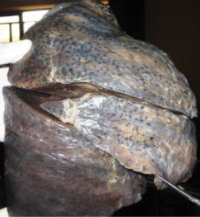 | |
| Fig. 1: | Right lung with complete oblique and horizontal fissure |
 | |
| Fig. 2: | Right lung with complete oblique fissure and incomplete horizontal fissure |
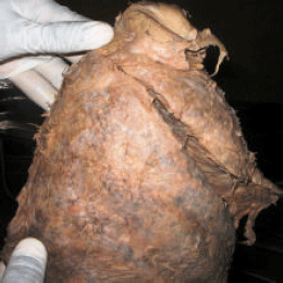 | |
| Fig. 3: | Right lung with oblique fissure and complete absence of horizontal fissure |
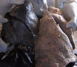 | |
| Fig. 4: | Right lung with complete horizontal and oblique fissure (left) and absence of horizontal fissure (right) |
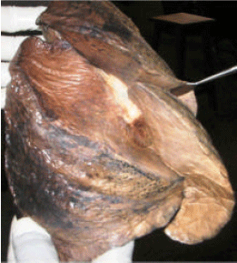 | |
| Fig. 5: | Right lung with complete oblique fissure and incomplete horizontal fissure |
OBSERVATIONS
Figure 1 showed a right lung with complete right and left fissures. We observed in Fig. 2 that the right lung showed a complete horizontal fissure and an incomplete oblique fissure. We also observed that Fig. 3 showed complete absence of horizontal fissure.
DISCUSSION
In prenatal life, fissures separate individual bronchopulmonary segments as the lungs grow. These fissures become obliterated except along two planes which is seen in fully developed lungs as oblique or horizontal fissures (Larsen, 1993). If pulmonary development is defective, it will give rise to variations in lobes and fissures of lung (Modgil et al., 2006). Absence or incomplete oblique or horizontal fissures could be due to obliteration of these fissures either completely or partially (Meenakshi et al., 2004)
The lobes of lungs show partial fusion as a result of incomplete pulmonary fissures. More than half of the pulmonary fissures are incomplete (Meenakshi et al., 2004). Studies abound on the variation in lung fissures (Medlar, 1947; Modgil et al., 2006). In Bates (1998), retrospective study of 1513 fetal postmortem examination reports 60 cases with anomalous fissures.
In another study of 1200 pairs of lungs, Medlar (1947), reported incomplete oblique fissure in 10.6 and 25.6% of the left-and right-sided lungs, respectively and incomplete horizontal fissure in 17.1% of the right-sided lungs. Oblique fissures were absent in 7.3% of the left-sided and 4.8% of the right-sided lungs; horizontal fissure was absent in 45.2% of the right-sided lungs. In Lukose et al. (1999) study on morphology of the lungs reported incomplete oblique fissure in 21% of left lung. Their study also revealed that the horizontal fissure was absent and incomplete in 10.5 and 21% of right lung, respectively. In Bergman et al. (1999) study on variations in peripheral segmentation of right lung and the base of the right and left lungs reported that the horizontal fissure was absent in 21% and incomplete in 67% of right lung. They also reported that 30% of right lung had incomplete oblique fissure. Furthermore, Meenakshi et al. (2004) reported that the horizontal fissure was absent in 16.6% and was incomplete in 63.3% of right lung while 46.6% of the left lung showed incomplete oblique fissure. From these studies, it can be deduced that absence of fissure is a common form of lung variation hence, the nature of fissure should be taken into consideration when planning operative strategy for thoracoscopic pulmonary resection where an incomplete fissure may contribute to post-operative air leakage (Meenakshi et al., 2004).
Furthermore, knowledge of fissure comes to bear when interpreting radiographic appearance of the lungs and during routine dissection of cadaver by students. In this study, we have presented variation in two right lungs as seen in the Gross Anatomy Laboratory of the College of Health Sciences, Niger Delta University (NDU), Bayelsa State, Nigeria and hope that this information will be useful in medical general medical practice.
REFERENCES
- Meenakshi, S., K.Y. Manjunath and V. Balasubramanyam, 2004. Morphological variations of the lung fissures and lobes. Indian J. Chest Dis. Allied Sci., 46: 179-182.
Direct Link - Modgil, V., S. Das, and R. Suri, 2006. Anomalous lobar pattern of right lung: A case report. Int. J. Morphol., 24: 5-6.
Direct Link - Bates, A.W., 1998. Variation in major pulmonary fissures: Incidence in fetal postmortem examinations and a review of significant extrapulmonary structural abnormalities in sixty cases. Pediatric Dev. Pathol., 1: 289-294.
PubMed - Lukose, R., P.S. Sunitha, M. Daniel, S.M. Abraham and M.E. Alex et al., 1999. Morphology of the lungs: Variations in the lobes and fissures. Biomedicine, 19: 227-232.
Direct Link